


Description
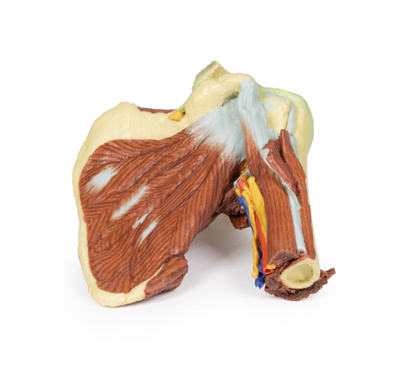
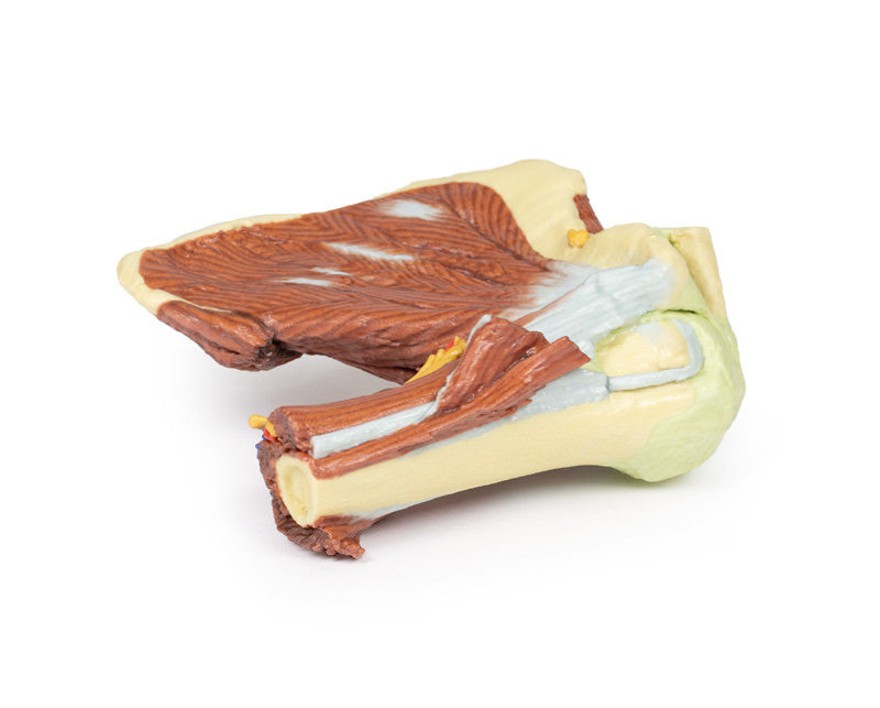
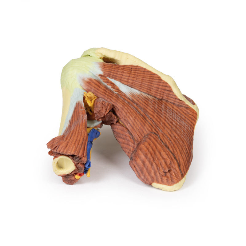
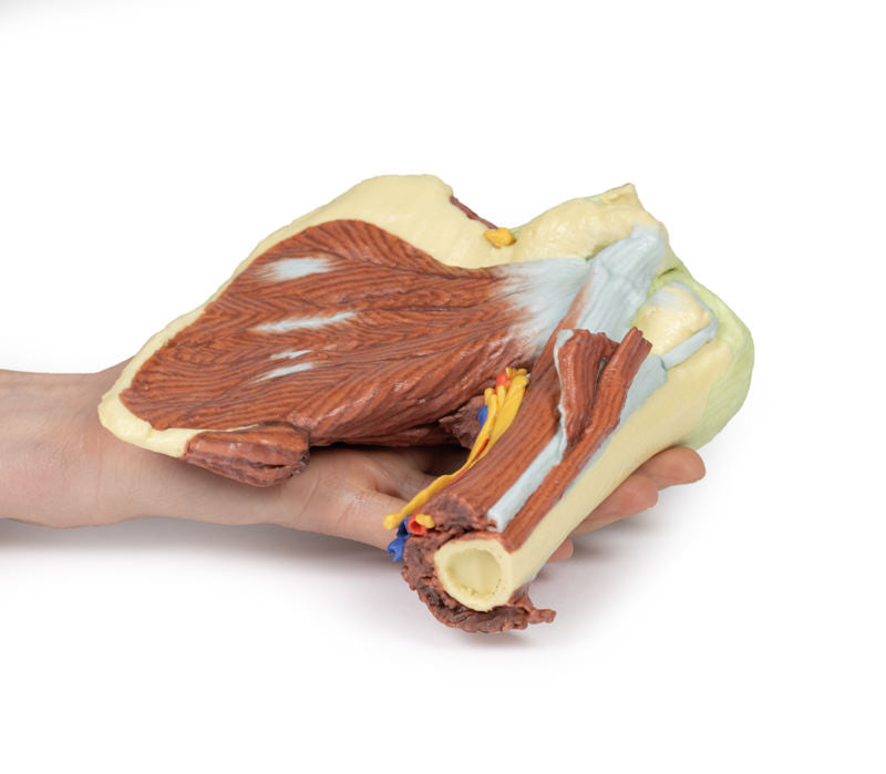
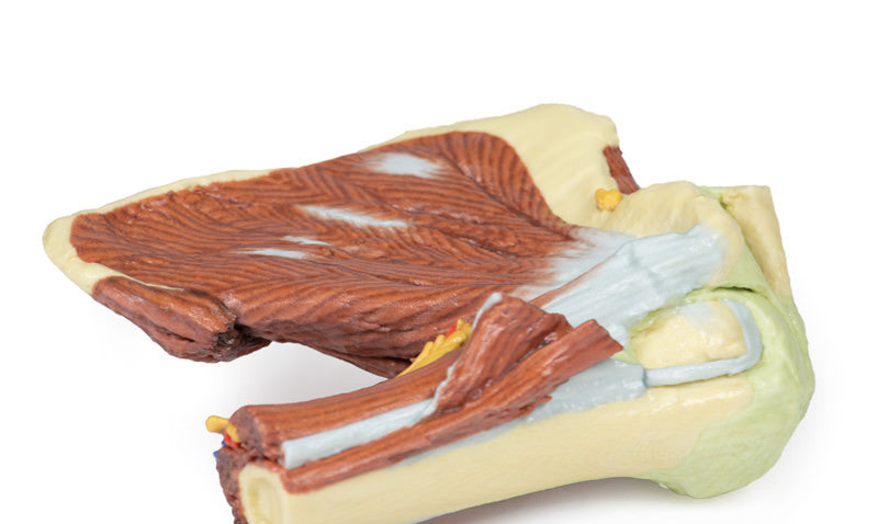
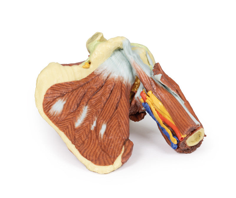
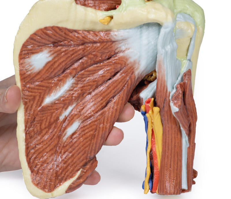
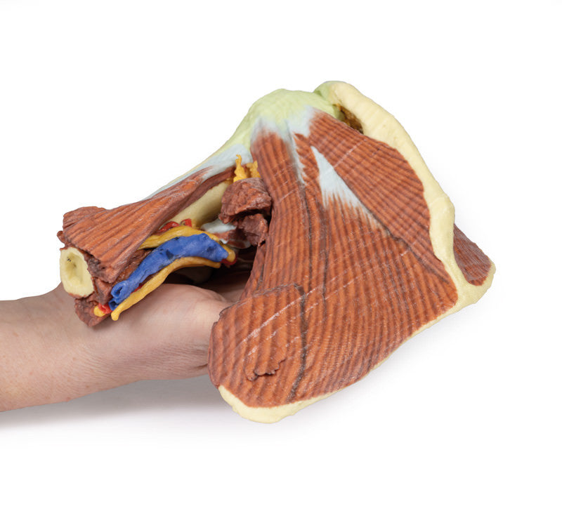
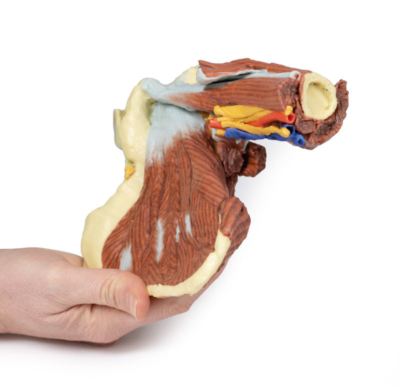
This 3D printed specimen presents a deep dissection of the left shoulder joint, musculature, and associated nerves and vessels of the scapula and proximal humerus (to near midshaft). Anteriorly, the deltoid muscle has been detached from its origin to expose the underlying deeper structures of the shoulder joint and rotator cuff musculature. The suprascapular nerve and artery are visible passing deep to, and superficial to, the superior transverse scapular ligament respectively. The multipennate subscapularis muscle is fully exposed with its tendinous insertion visible deep to the short head of the biceps brachii muscle. The insertion of the deltoid is preserved just overlying the long head of the biceps brachii, which ascends through the bicipital groove towards the glenohumeral joint capsule. Adjacent to the short head of the biceps brachii is the neurovascular bundle of the brachial artery, brachial vein, and terminal nerves of the brachial plexus (radial, ulnar, median, and the medial antebrachial cutaneous). The tendon of the latissimus dorsi, teres major, teres minor and long head of the triceps brachii muscles have been cut enhance the visibility of the medial aspect of the humerus, including the passage of the axillary nerve into the quadrangular space, the origin of the profunda brachii artery accompanying the radial nerve, and the insertion of the short head of the triceps brachii muscle.
On the posterior aspect, the infraspinatus and supraspinatus muscles are fully exposed from their origins to insertions on the proximal humerus. The glenohumeral joint capsule is intact, with the extracapsular ligaments (e.g., acromioclavicular, coracoacromial, and coracoclavicular [both conoid and trapezoid portions]) preserved.
Please note that all of these items are produced upon order and do require roughly 4 - 6 weeks for delivery. All items are produced in Germany and do take some time for transport. We will provide updates on delivery timeframes upon order.
