


Description
Clinical History
A 38-year-old female presents with severe nausea, vomiting, fevers, and rigors. She has a history of recurrent urinary tract infections over the past 6 months, requiring several courses of oral antibiotics and one admission for IV antibiotics. Blood tests show raised inflammatory markers. Urinalysis is positive for white blood cells (WBC). A CT scan reveals unilateral left hydronephrosis and pyelonephritis. She fails to respond to conservative treatment and undergoes a nephrectomy. She makes a complete recovery.
Pathology
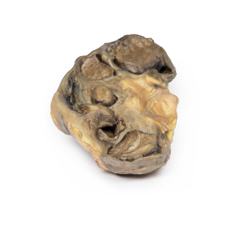
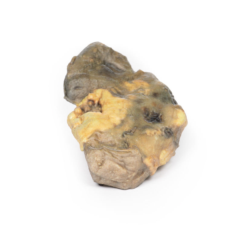
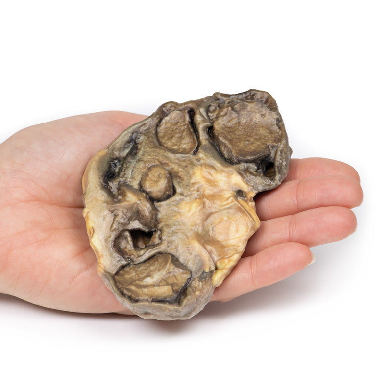
The patient’s left nephrectomy specimen shows a kidney sliced to display the cut surface. The pelvis and calyces are greatly dilated and contain remnants of yellow pus. There is considerable fibrosis of the renal parenchyma. Near the mid-zone lateral border, there is a hemorrhagic necrotic area measuring 35 x 12 mm containing pus. Two smaller hemorrhagic necrotic areas are visible on the capsular surface. These lesions likely represent hemorrhage into an abscess cavity and are continuous with the lesion seen on the cut surface, consistent with a perinephric abscess.
Further Information
Pyonephrosis occurs when obstruction in the upper urinary tract combines with pyelonephritis. Debris from infection, WBCs, and bacteria accumulate in the obstructed kidney, leading to a hydronephrotic kidney filled with pus. Staghorn calculi often form in chronic or recurrent infections due to an alkaline urinary pH caused by bacteria.
Pyonephrosis is rare. Risk factors include immunosuppression, diabetes, and anatomical urinary tract obstructions such as strictures, horseshoe kidneys, tumours, and urinary calculi.
Clinical presentation may be vague but typically includes constitutional symptoms of sepsis, flank pain, haematuria, dysuria, and pyuria. A grossly enlarged kidney may be palpable. Pyuria is present on urinalysis.
Diagnosis is primarily radiological, using CT, ultrasound, or MRI to identify obstruction and pyelonephritis.
Treatment depends on the underlying cause of obstruction. Emergent management involves drainage of pus, performed via percutaneous or retrograde ureteral stents by urology or interventional radiology. Surgical treatment depends on the obstruction’s cause. Antibiotics are essential for infection control.
If untreated, complications include florid sepsis, xanthogranulomatous pyelonephritis, renal or perinephric abscess formation, and fistulas to pleura, colon, or duodenum.
