


Description
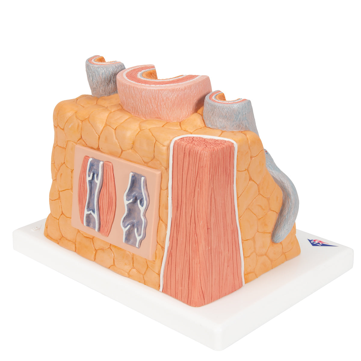
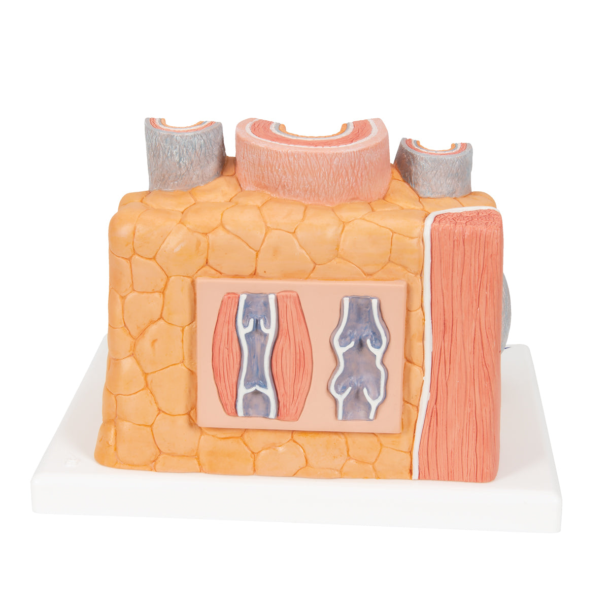
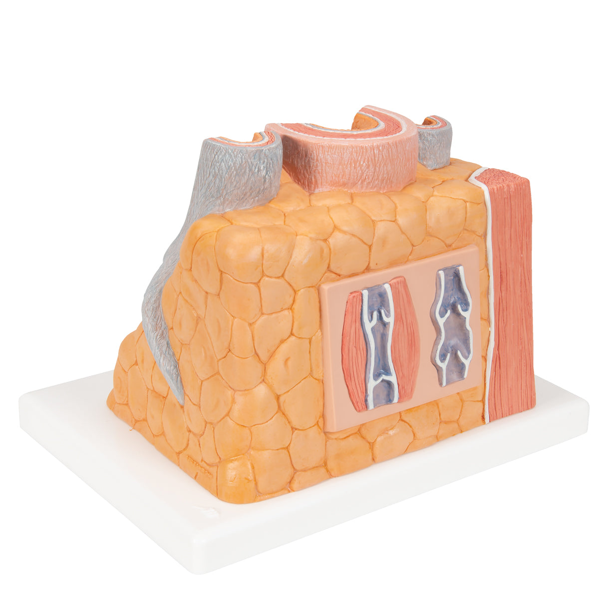
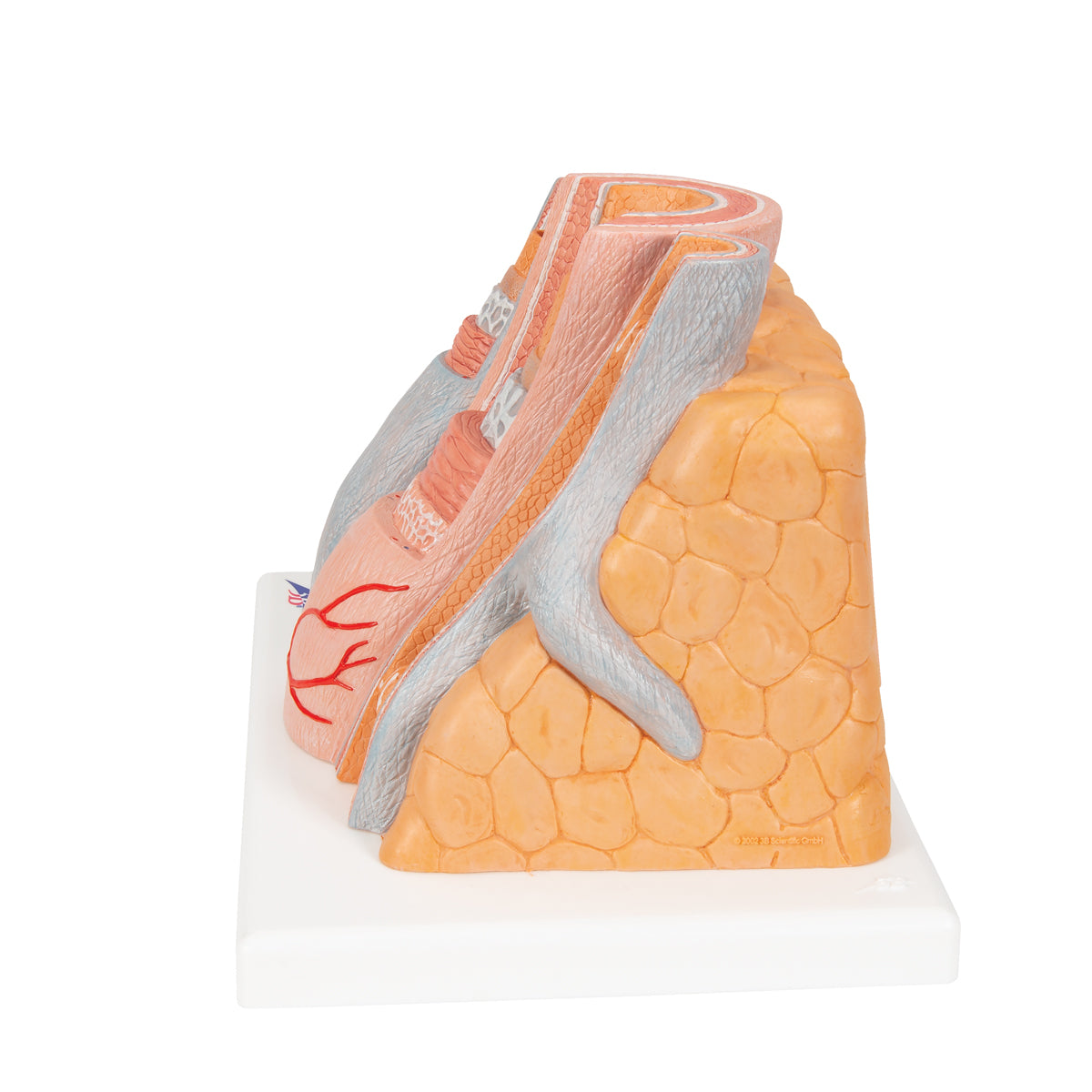
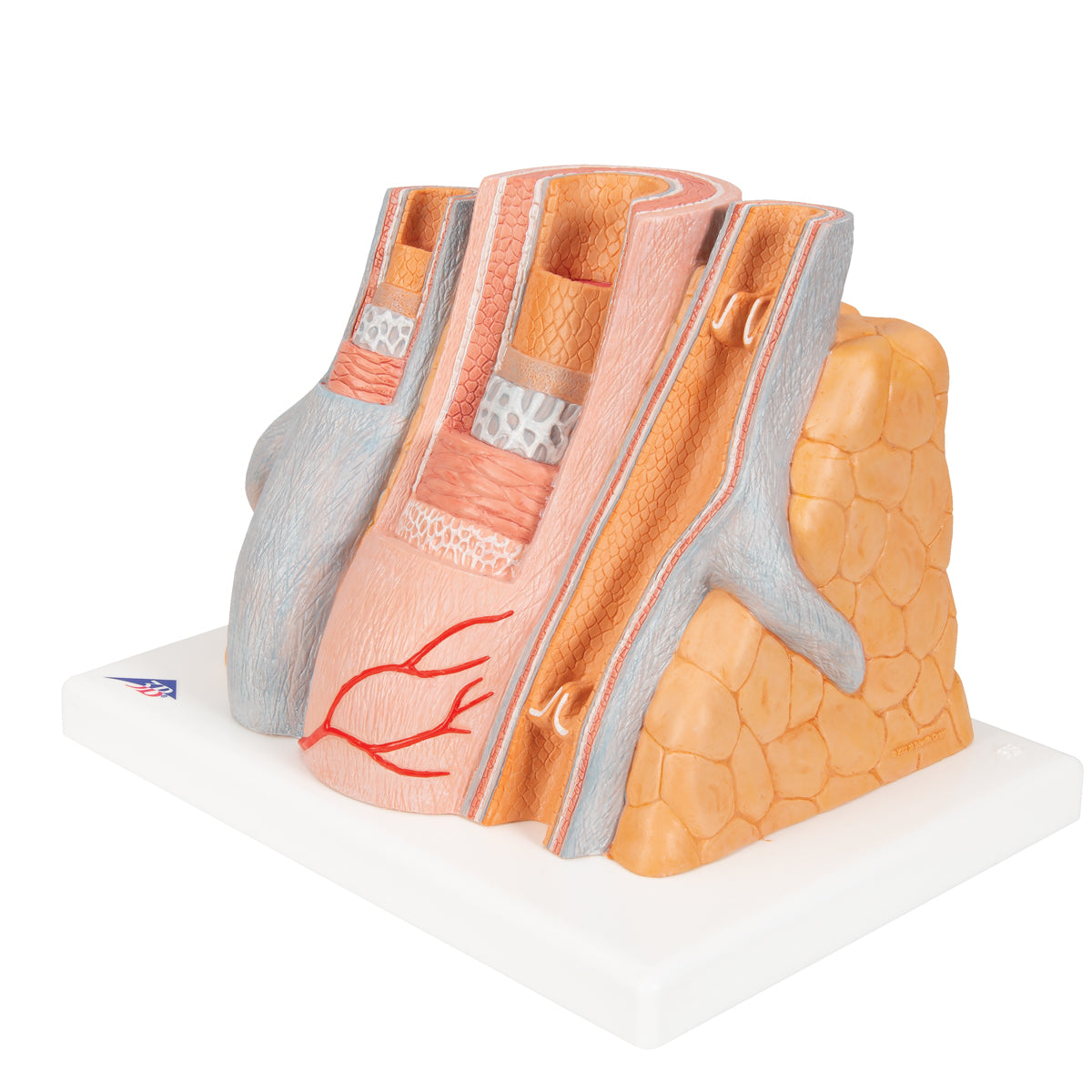
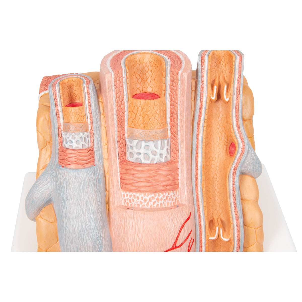
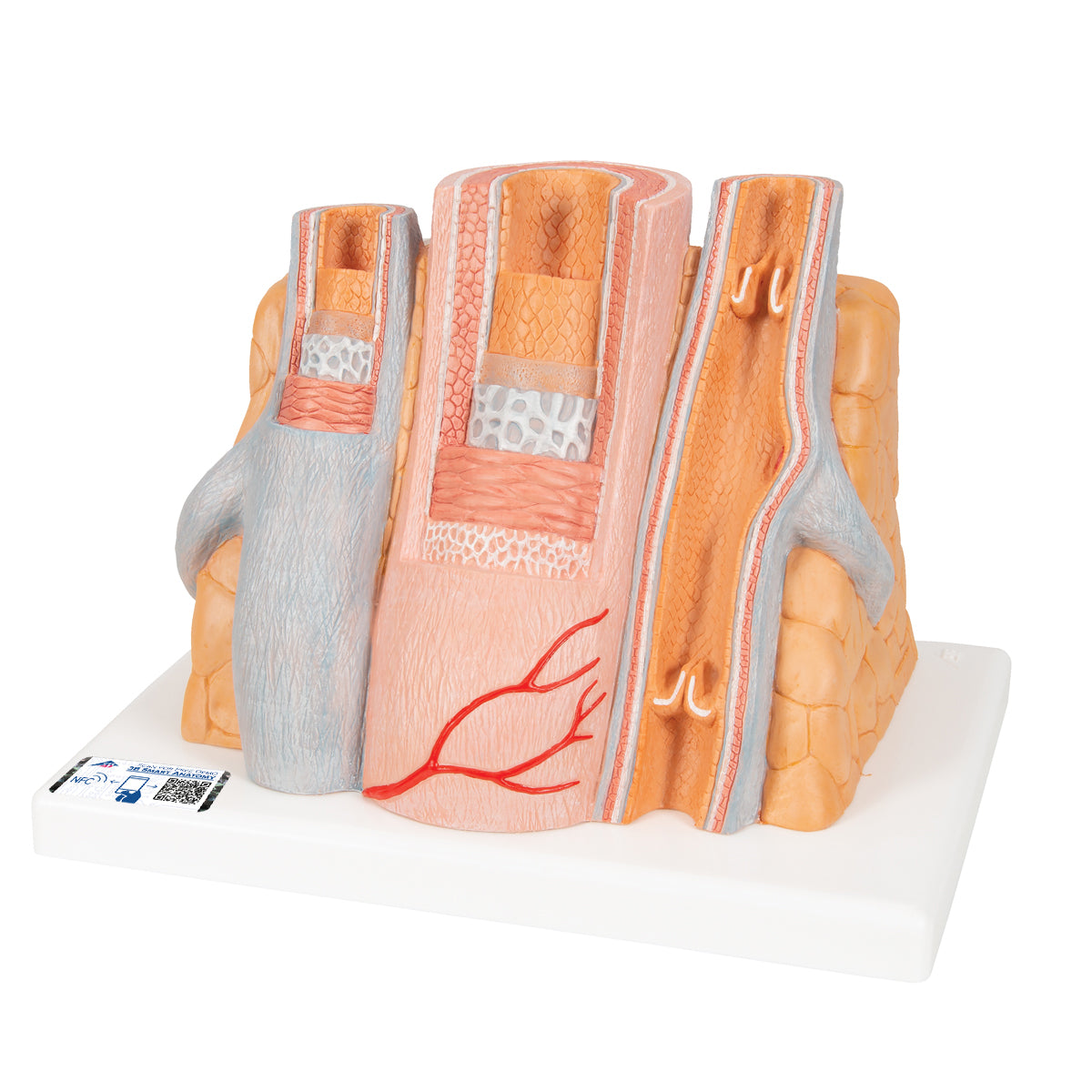
Vascular anatomy and histology programs studying the microscopic structure of blood vessels benefit from highly magnified models, and this 14-times enlarged 3B Scientific artery and vein model presents muscular artery and adjacent vein wall anatomy in precise detail.
The artery and vein model shows a medium-sized muscular artery with two adjacent veins from the antebrachial area with adjoining fat tissue and muscle enlarged 14 times. The MICROanatomy circulatory system model illustrates the reciprocal anatomical relationship of artery and vein and the basic functional techniques of the venous valves (“valve function” and “muscle pump”). The left vein and the middle artery are fenestrated in the upper anterior segment, revealing the various layers of the wall structure in a cross and longitudinal section and in top view. The right vein is opened throughout in the anterior segment, revealing the orifice of a feeder vein and two venous valves, i.e. “flap valves” formed by a duplication of the tunica intima. On the rear of the artery and vein model, the relief of two veins is shown to illustrate the functional aspect of the venous valves. A great tool for teaching about the human circulatory system. Artery and vein supplied on base.This product features 3B Smart Anatomy for easy access to model information — read more below.
Specifications
| Weight | 0.97 kg (2.1 lbs) |
| Dimensions | 26 x 19 x 18.5 cm (10-1/4" x 7-1/2" x 7-1/4") |
| Warranty | 5 year |
This item is enhanced with SmartAnatomy!
The new way to experience and supplment your model with online, interactive content. No more reference guides and booklets - access product details right from your mobile device. Learn more

3B Scientific Authorized Distributor

Produced by the 3B Scientific, leaders of high quality healthcare and scientific models. Learn about their story.
Products are backed by the manufacturer warranty.