


Description
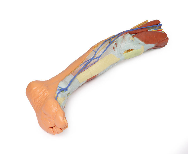
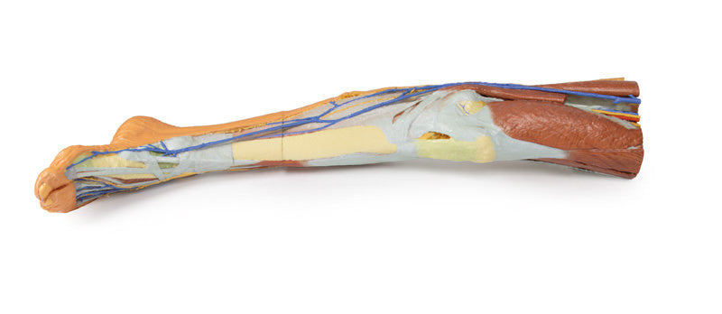
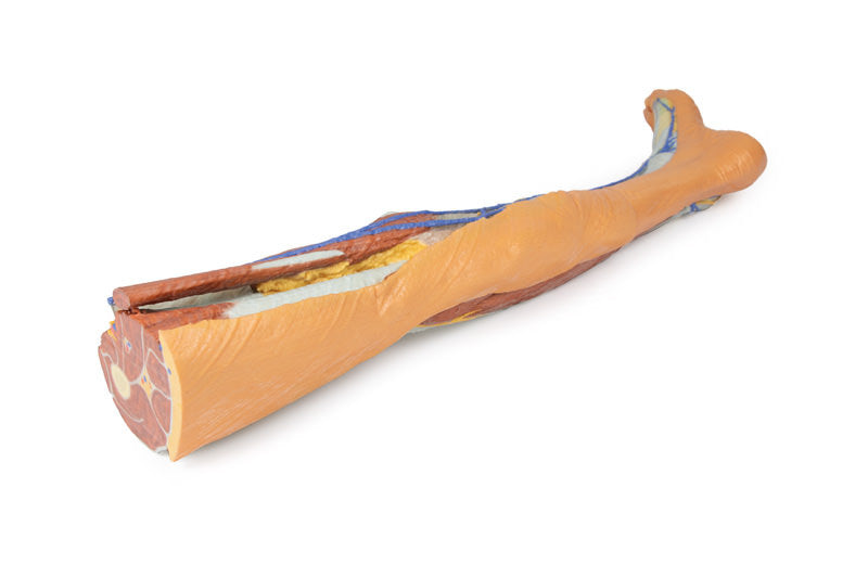
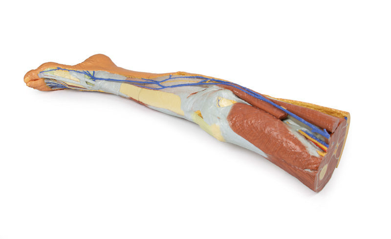
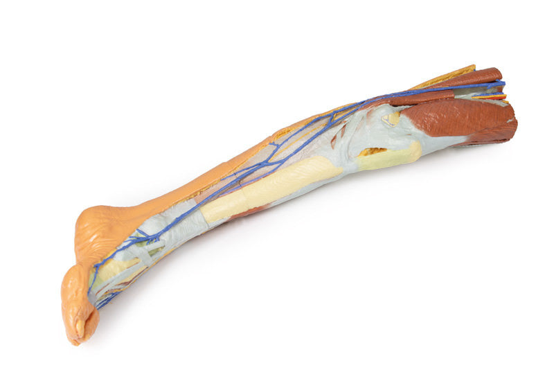
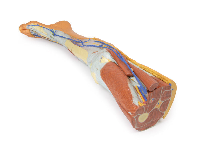
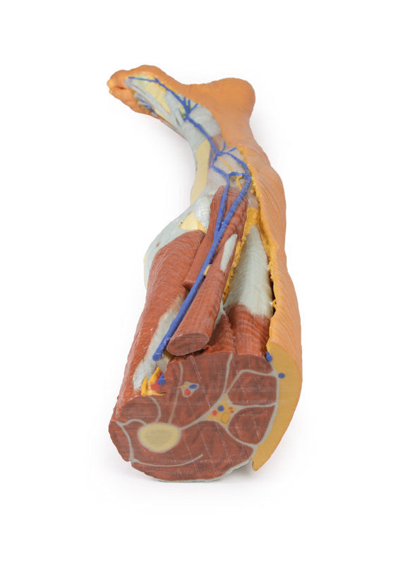
This 3D printed specimen represents the remainder of the lower limb portions of our male abdominopelvic and proximal thigh specimen (MP1765), sectioned proximally near midthigh and continuous to the partially dissected foot. The transverse section through the thigh exposes the neurovascular structures of the anterior, medial and posterior compartments. This includes the great saphenous vein superficial to the terminal branches of the femoral nerve, femoral artery and vein in the anterior compartment, and perforating branches of the deep femoral artery in the medial and posterior compartments.
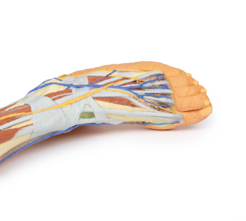
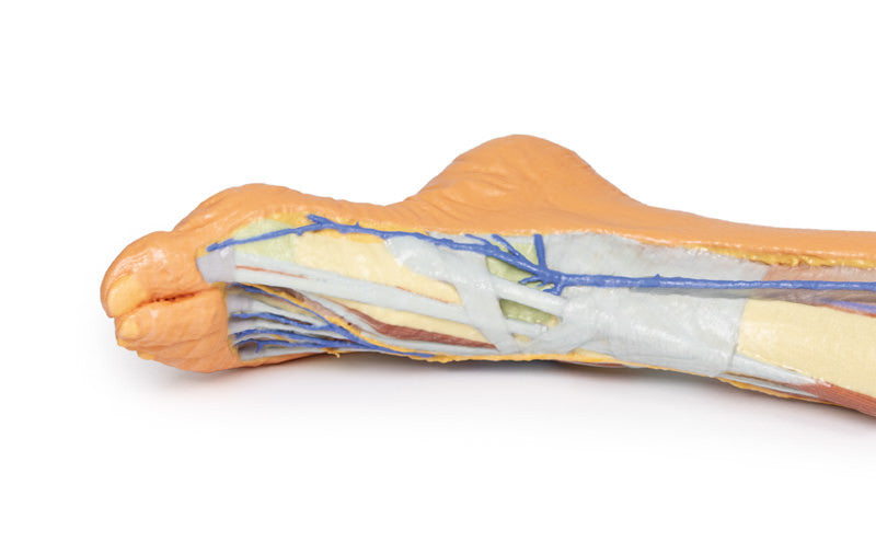
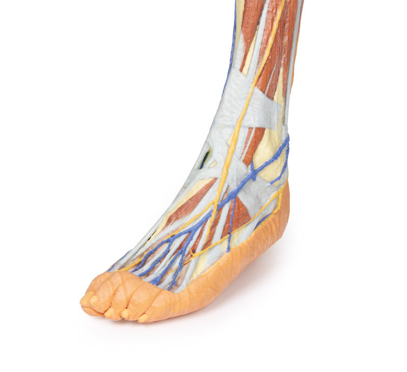
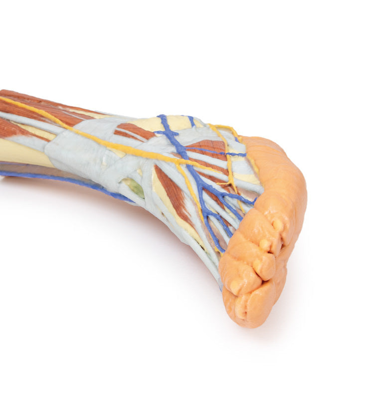
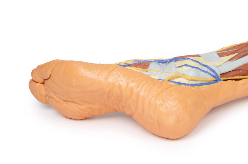
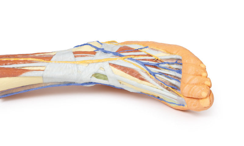
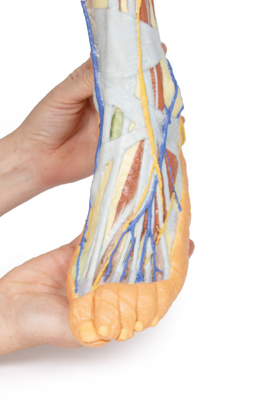
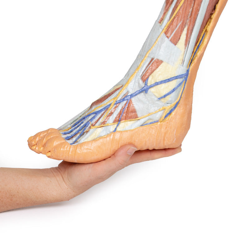
The remainder of the thigh, leg and dorsum of the foot have been dissected to demonstrate superficial structures and compartmental musculature, except the posterior aspect of the specimen which has been left undissected. The course of the great saphenous vein is displayed from the medial aspect of the thigh to the medial malleolus and the medial aspect of the dorsal venous plexus. The origin of the small saphenous vein from lateral branches of the dorsal venous plexus is also visible to the margin of the dissected superficial fascia near the lateral malleolus. The deeper femoral artery, vein and nerve branches are visible deep to the anterior compartment musculature (and a sectioned sartorius muscle) entering the adductor canal. Near the medial aspect of the knee joint the saphenous nerve is visible passing superficially near the great saphenous vein on the surface of the posterior crural fascia and terminating as the medial cutaneous nerve of the leg branches. On the lateral aspect of the leg the medial and intermediate dorsal cutaneous branches from the superficial fibular nerve are preserved passing onto the dorsum of the foot adjacent to the dorsal venous plexus tributaries.
Please note that all of these items are printed upon order and do require roughly 4 - 6 weeks for delivery. All items are produced in Germany. We will provide updates on delivery timeframes upon order. Please note that once placed and processed, an order cannot be cancelled or altered.
Erler-Zimmer Authorized Distributor

Produced by Erler Zimmer. Learn about their story.
Products are backed by a 3 year warranty.