


Description
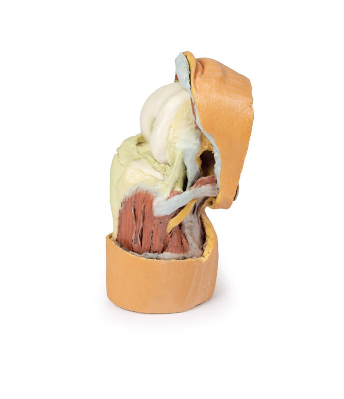
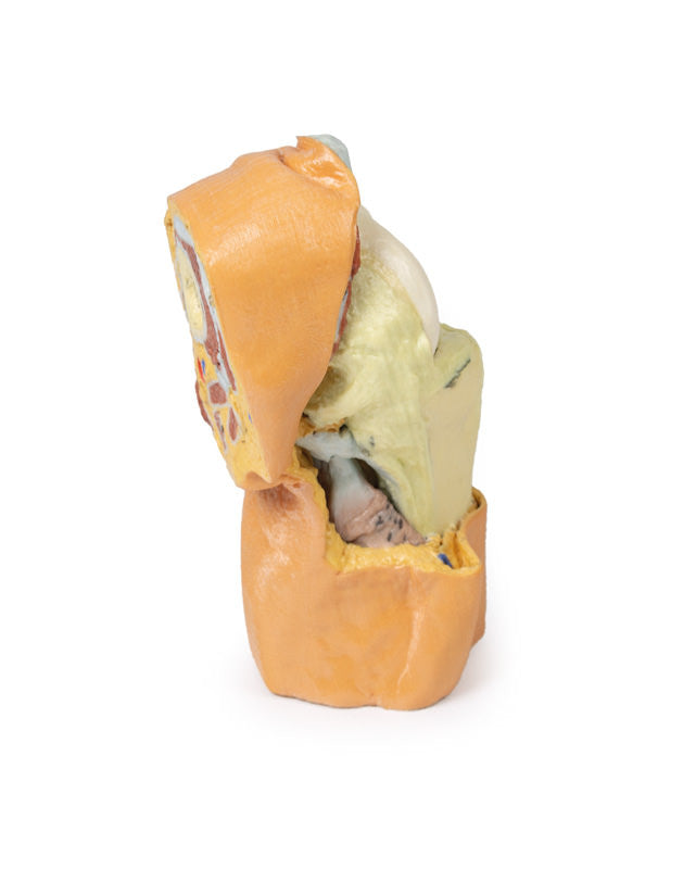
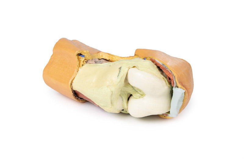
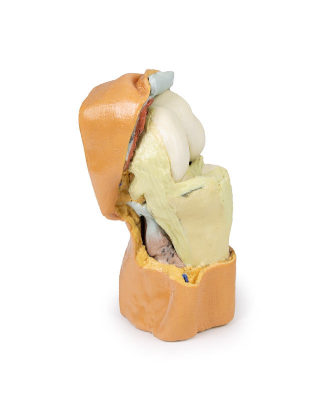
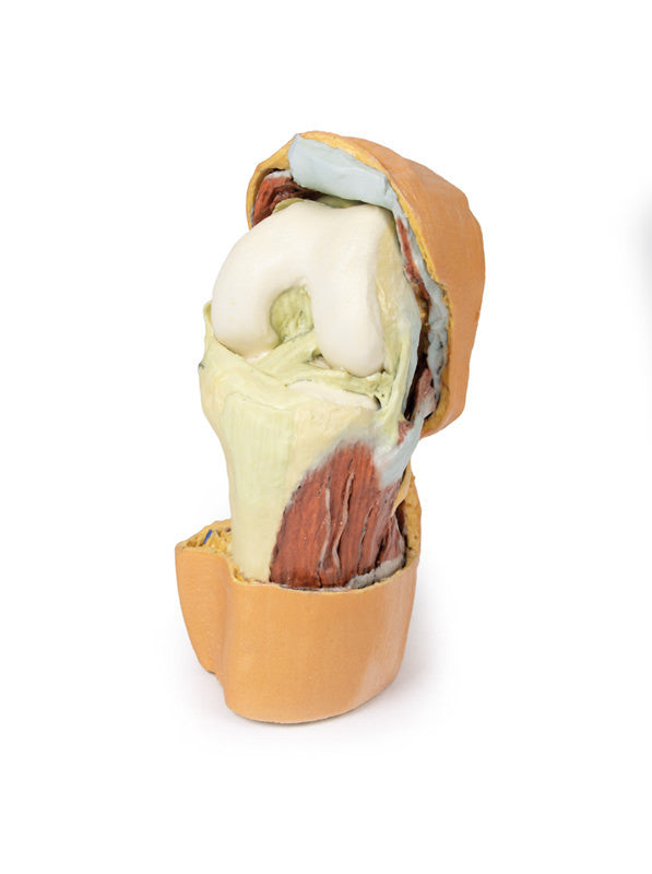
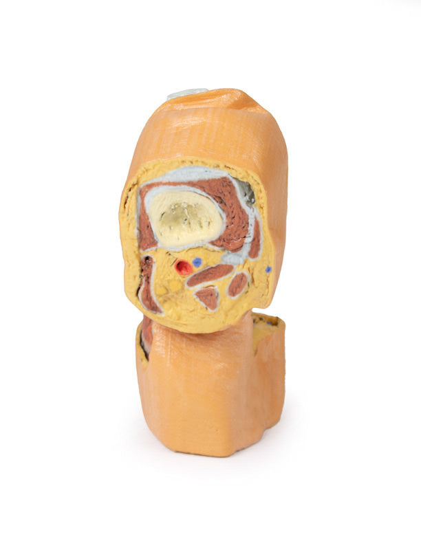
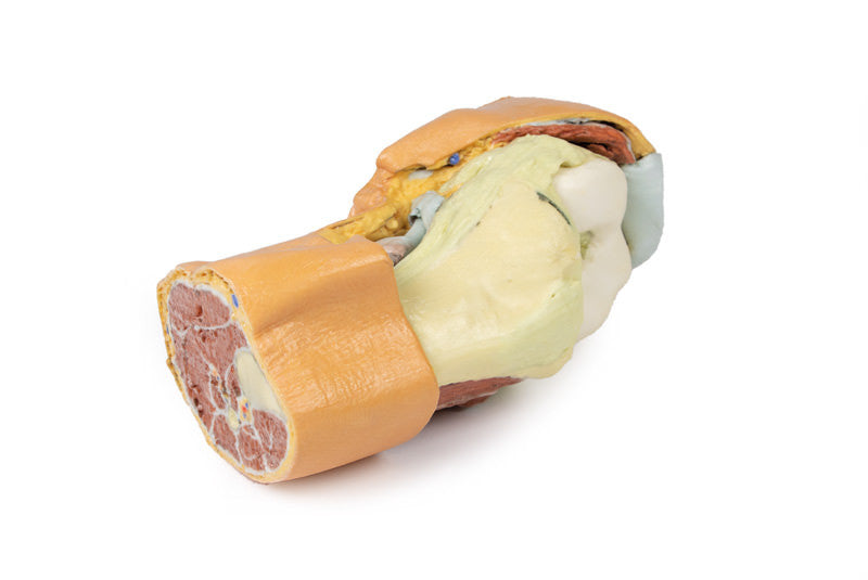
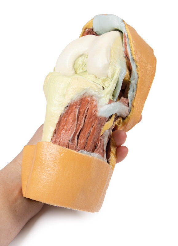
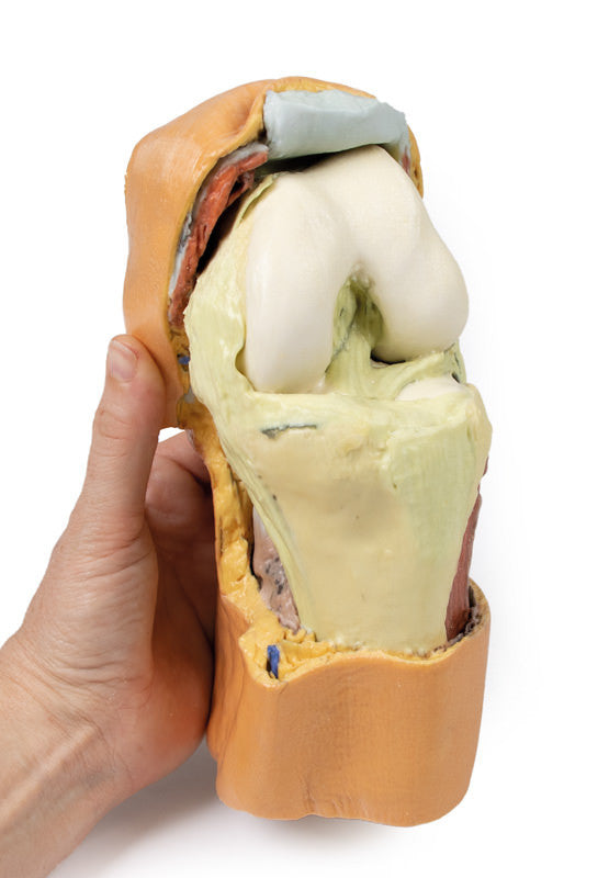
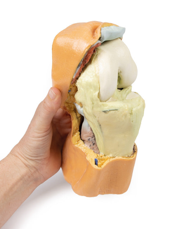
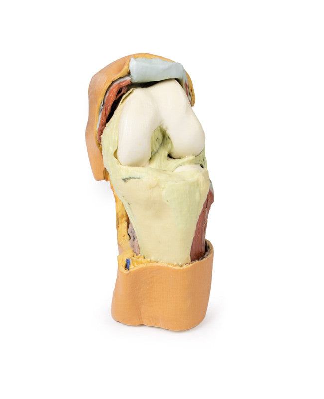
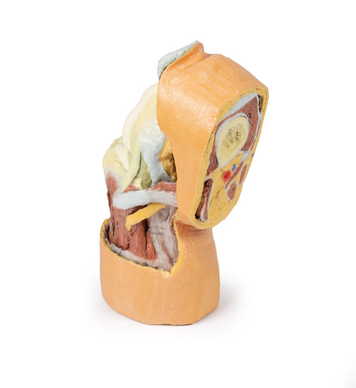
This 3D printed specimen displays a deep dissection of a left knee joint with the internal joint capsule structures relative to superficial tissues in a flexed position. The proximal cross-section through the distal thigh captures a small portion of the quadriceps femoris and sartorius anteriorly (with the thickened connective tissue of the iliotibial tract), the fat-filled popliteal fossa (with the popliteal vessels, tibial and common peroneal nerves), and the termination of the medial (adductor magnus tendon, gracilis) and posterior thigh muscles (biceps femoris, semitendinosus, semimembranosus) posteriorly. The distal cross-section through the leg preserves the most proximal portion of the muscles of the anterior, lateral and posterior compartments. Also visible in the section are the associated neurovascular structures: the anterior tibial artery, vein and deep peroneal nerve; the posterior tibial artery, vein and tibial nerve; and the fibular artery and vein.
Anteriorly, the skin, subcutaneous tissue and patella have been removed, with only remnant portions of the tendon of the quadriceps femoris and patellar ligament retained. With the joint capsule opened, the anterior and posterior cruciate ligaments and the menisci are visible positioned between the femoral condyles and tibial plateau. On the medial aspect, the tibial (medial) collateral ligament passes just anterior to the insertion of the semitendinosus of the pes anserinus (the sartorius and gracilis tendons are sectioned), which in turn is just anterior to the posterior compartment musculature (covered by crural fascia). On the lateral aspect, the fibular (lateral) collateral ligament is preserved, and the anterior crural musculature is exposed. Passing from the thigh are the inserting tendon of the biceps femoris onto the head of the fibula, as well as the common peroneal nerve.
Please note that all of these items are printed upon order and do require roughly 4 - 6 weeks for delivery. All items are produced in Germany. We will provide updates on delivery timeframes upon order. Please note that once placed and processed, an order cannot be cancelled or altered.
