


Description
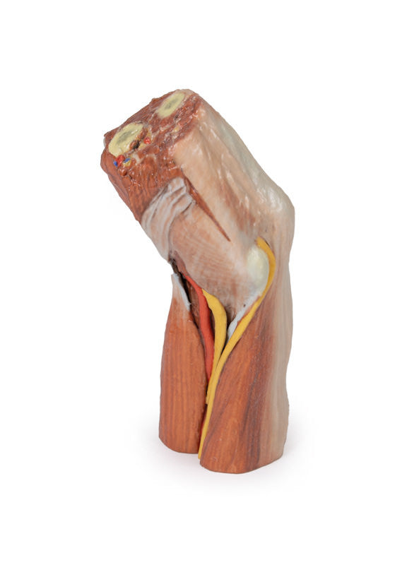
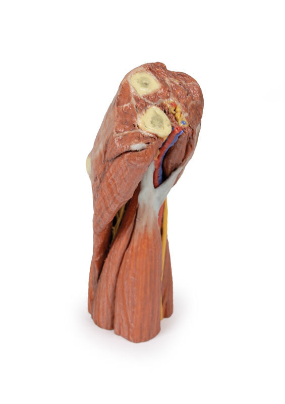
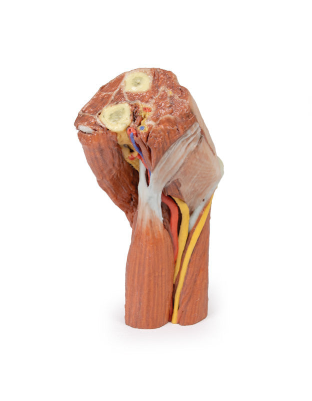
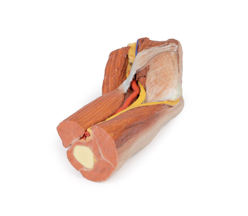
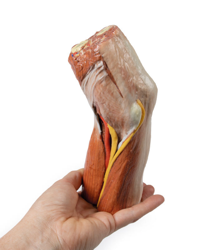
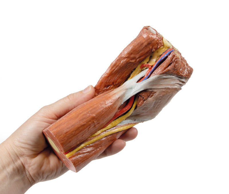
This 3D printed specimen presents a left distal arm and proximal forearm with all skin, subcutaneous fat and superficial cutaneous nerves and veins removed. The elbow region partially flexed to display the arrangement of muscles and neurovascular structures of the cubital fossa.
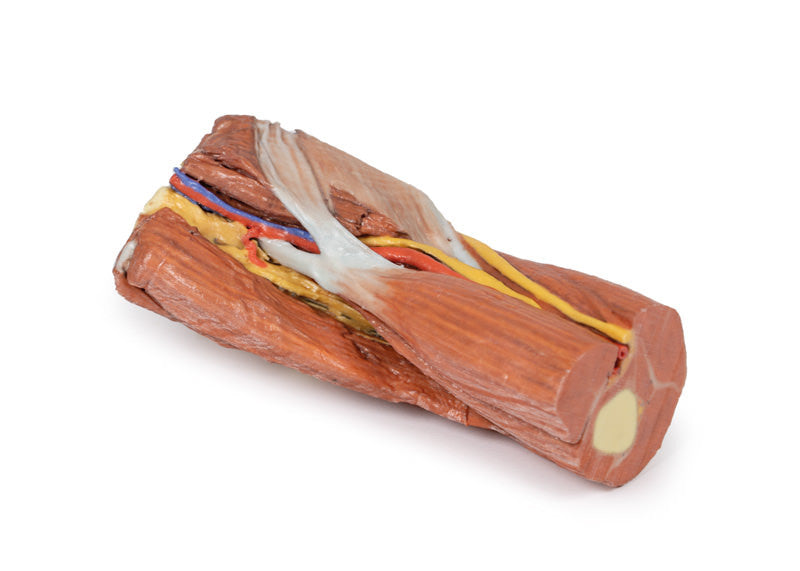
Viewed from the anterior aspect the most obvious feature is the biceps brachii muscle, with its insertion in the form of the flattened bicipital aponeurosis passing medially over the muscles of the common flexor origin and the more rounded tendon passing deep to insert into the radial tuberosity. The brachialis muscle lies deep to biceps brachii and is visible from the lateral aspect. In the proximal part of the forearm the brachioradialis muscle (slightly elevated and reflected laterally to reveal deeper structures) and extensor carpi radialis longus are identifiable. On the medial side one can see the classic arrangement of the biceps brachii tendon, brachial artery and median nerve (TAN) from lateral to medial. They are partially covered by the bicipital aponeurosis as they course distally. The ulnar nerve can be seen changing position from the anterior compartment of the arm to the posterior compartment (the intermuscular septum has not been preserved but triceps muscle is clearly evident) to pass behind the medial epicondyle and enter the cubital tunnel. It travels distally between the two heads of flexor carpi ulnaris. Close inspection of the groove between brachialis and brachioradialis reveals the radial nerve (which would not be visible if the brachioradialis muscle had not been partly reflected). It lies amongst some fat (yellow) but its superficial branch passes distally below brachioradialis.
In the posterior view the triceps tendon inserts into the olecranon process of the ulna. The medial and lateral epicondyles are also clearly visible (grey/white in colouration). The medial epicondyle is clearly identifiable as it has the ulnar nerve passing posteriorly before penetrating the deep fascia covering the gap between the two heads of flexor carpi ulnaris.
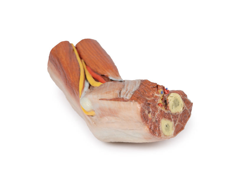
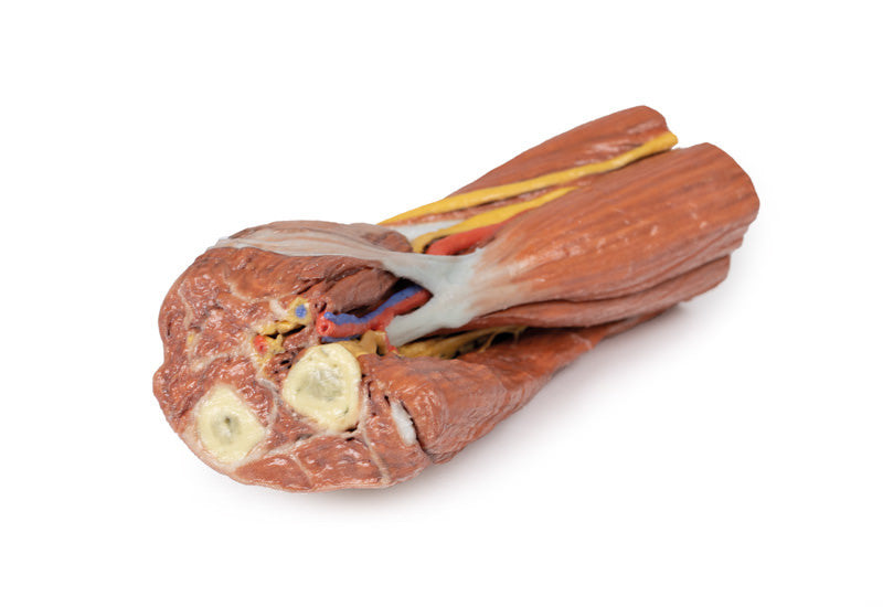
The proximal section through the arm reveals the biceps muscle lying anteriorly with the neurovascular bundle on its medial side which contains the brachial artery together with median nerve and ulnar nerve (veins have been removed). The three heads of triceps (lateral, long and the deeper placed medial head) are clearly visible in the posterior compartment. On the distal section through the forearm it is more difficult to discern each muscle, but the cut surfaces of the radius and ulna are clearly visible - as is the brachial artery lying medial to pronator teres muscle and the median nerve lying just deep to this muscle (which is the most lateral of the muscles arising from the common flexor origin).
Please note that all of these items are printed upon order and do require roughly 4 - 6 weeks for delivery. All items are produced in Germany. We will provide updates on delivery timeframes upon order. Please note that once placed and processed, an order cannot be cancelled or altered.
Erler-Zimmer Authorized Distributor

Produced by Erler Zimmer. Learn about their story.
Products are backed by a 3 year warranty.