


Description
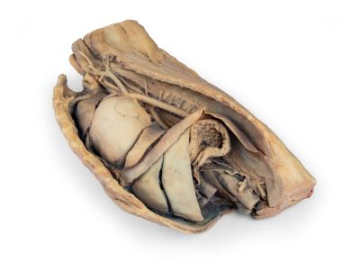
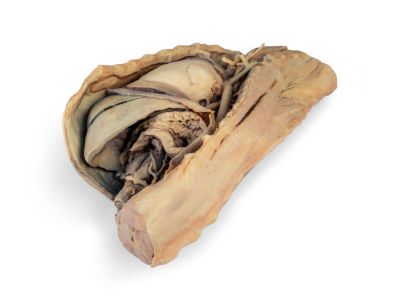
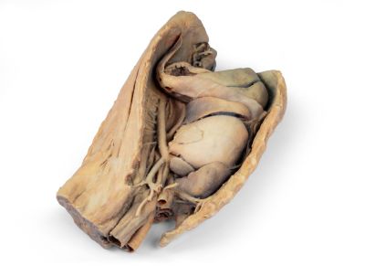
This specimen depicts the topography of the heart following the removal of the left lung during a dissection of the thoracic cavity and cranial abdomen of a dog approached from the left side. The cranial and accessory lobes of the right lung remain in situ, serving as anatomical reference points. Within the cranial mediastinum, the brachiocephalic trunk and the left subclavian artery are identifiable as major branches arising from the aortic arch.
The thoracic portion of the esophagus runs through the mediastinum in a craniocaudal direction. The diaphragm has been preserved to serve as a landmark for retrodiaphragmatic organs such as the liver and the stomach.
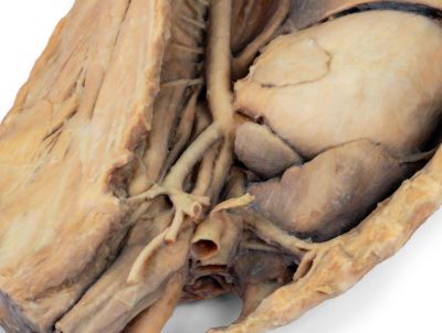
The diaphragmatic crura and their relation to the aortic hiatus are maintained, as are the initial visceral branches of the abdominal aorta—specifically, the celiac trunk and the cranial mesenteric artery.
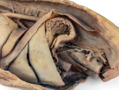
Both the interventricular and coronary grooves are visible. A portion of the right atrial wall has been removed to expose the right atrioventricular (tricuspid) valve, including the chordae tendineae and papillary muscles.
Additionally, the septomarginal trabecula (moderator band) is visible within the right ventricle, as well as the trajectory from the ventricle to the pulmonary trunk. The left atrial wall is also open to display its lumen.
