Canadian veterinary education programs demand training tools that deliver authentic clinical experiences while accommodating the practical realities of academic instruction. The Erler Zimmer Lillie Canine Ultrasound Phantom Training Kit (VET4850) addresses these needs by providing students with realistic, repeatable ultrasound training that builds competency before clinical rotations, while offering faculty a reliable, low-maintenance teaching resource.

Building Clinical Confidence Through Realistic Training
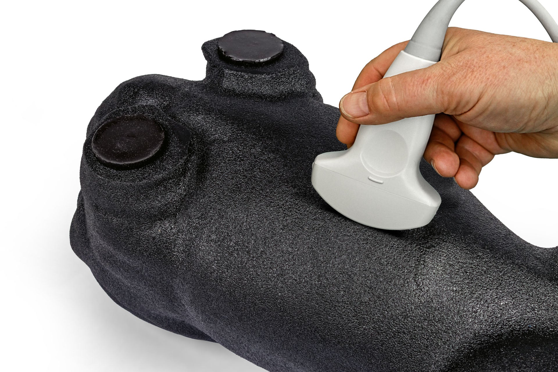
For students entering veterinary ultrasound, the transition from theory to live-animal scanning presents significant challenges. Lillie bridges this gap by offering a tissue-mimicking training platform that allows learners to develop foundational scanning skills in a controlled, pressure-free environment. Students can practice probe manipulation, optimize machine settings, and refine their technique through unlimited repetitions—building the muscle memory and spatial awareness essential for confident clinical performance.
Each training kit arrives in a robust, lockable case designed for both secure storage in simulation labs and easy transport between teaching locations. This portability allows programs to maximize their investment by enabling demonstrations in multiple learning environments, from traditional classrooms to clinical skills labs.
Quality Assurance for Academic Programs
Understanding the importance of consistency in educational tools, every Lillie phantom undergoes individual ultrasound inspection before shipment. This quality control process includes documentation of all features and characteristics, ensuring your program receives a training tool that performs predictably across multiple student cohorts. The specialized tissue-mimicking plastic construction provides durability for extended academic use, and included care instructions help maintain optimal phantom condition throughout years of student training sessions.
Comprehensive Anatomical Training Across Multiple Positions
Handcrafted through a meticulous 70+ step production process utilizing 35 specialized moulds, each Lillie phantom includes key canine abdominal structures designed to provide authentic ultrasound imaging characteristics. While not an exact anatomical replica, the phantom's detailed construction closely mimics real patient imaging, giving students meaningful preparation for clinical work.
Position #1: Dorsal Recumbency
In dorsal recumbency, students develop foundational scanning skills by identifying essential abdominal structures including the stomach, pyloric region, duodenum, liver, and gallbladder in both longitudinal and transverse planes. This position emphasizes mastery of machine controls—depth, gain, time gain compensation (TGC), dynamic range, and zoom—allowing learners to understand how these adjustments affect image quality and diagnostic clarity. Students refine probe positioning through systematic rotation and sliding techniques, capturing structures in multiple orientations to build spatial understanding. Measurement exercises, such as assessing the cystic duct, develop the precision required for clinical documentation. Faculty can use this position to establish reproducible scanning protocols that students will carry forward into live-animal practice.

Position #2: Left Lateral Recumbency
Left lateral recumbency training focuses on localizing and evaluating the right kidney and adrenal gland, advancing students' skills in optimizing focus depth, focal zones, frequency selection, and zoom for detailed anatomical imaging. This position emphasizes measurement accuracy and repeatability—critical skills for monitoring disease progression or treatment response in clinical practice. Students follow the duodenum's path in both planes, isolating probe movements to maintain image optimization while tracking anatomical structures. Identification and imaging of the right pancreatic limb further develops anatomical recognition and scanning versatility, preparing students for the subtle probe adjustments required in clinical ultrasonography.

Position #3: Right Lateral Recumbency
In right lateral recumbency, students concentrate on imaging the left kidney in both longitudinal and transverse views, practicing the probe alignment techniques necessary to maintain proper organ orientation for accurate measurements. Comparative measurement exercises—with the kidney positioned horizontally versus non-horizontally—demonstrate the significant impact of probe positioning on diagnostic accuracy. Training extends to the left adrenal gland, where students adjust depth, frequency, zoom, and focus to achieve clear, reproducible assessments. Students also examine the spleen's extent and echotexture, practicing the overlapping sweep technique essential for comprehensive organ evaluation. Urinary bladder scanning in multiple planes reinforces students' ability to optimize controls for highlighting anechoic content and achieving clear organ margins—fundamental skills for abdominal ultrasonography.

Systematic Ultrasound Assessment Framework
To support standardized teaching and assessment, the Lillie training program incorporates the LEMONS framework—a systematic approach to ultrasound evaluation that students can apply consistently across all structures:
- Location (anatomical position, focal vs. diffuse distribution)
- Echotexture (hypoechoic, isoechoic, hyperechoic, heterogeneous, homogeneous)
- Measurements (qualitative or quantitative assessment in mm or cm)
- Outline (margination quality, regularity, definition)
- Numbers (quantifying lesions or structures)
- Shape (morphological description: ovoid, rounded, fusiform, amorphous)
This structured approach gives faculty a consistent framework for teaching and evaluating student competency while preparing students for professional reporting standards used in clinical practice.
Advanced Skills Development with the Training Block
The included skills phantom block provides faculty with an excellent tool for teaching three-dimensional spatial reasoning—one of ultrasound's most challenging concepts for students to grasp. By scanning embedded objects from multiple angles, students build mental maps of complex three-dimensional shapes, a skill directly transferable to clinical interpretation of patient anatomy.
The block facilitates progressive skill development as students track anechoic tubular structures in multiple planes, developing the fine motor control required for following vessels, ducts, and other tubular anatomies in live patients. Accurate luminal diameter measurement with proper caliper placement reinforces measurement precision that students will apply throughout their clinical careers.

Fine Needle Aspiration Training
The self-healing fine needle aspiration (FNA) training component allows students to develop procedural competency in ultrasound-guided sampling techniques before performing these procedures on patients. Students practice the complete workflow: assessing target structures using the LEMONS criteria, planning their approach, adjusting dynamic range to enhance needle visibility, and executing the procedure with proper technique.
The training emphasizes practical skills including probe stabilization, hand-position selection for optimal control, and in-plane needle guidance toward the target. Ambidextrous training—switching probe and needle hands—ensures students develop versatility for various clinical scenarios and patient positioning requirements. While the self-healing material withstands repeated use, faculty should note that intensive practice sessions may eventually produce needle marks that affect block longevity—a worthwhile trade-off for thorough procedural training.
Supporting Canadian Veterinary Education
As the Canadian authorized distributor for the Lillie Canine Ultrasound Phantom, we're committed to supporting veterinary and simulation programs across Canada in delivering exceptional ultrasound education. The combination of realistic anatomical representation, durable construction, and comprehensive training applications makes Lillie an invaluable addition to any veterinary curriculum.
For programs seeking to enhance their ultrasound training capabilities or to discuss how Lillie can meet your specific educational objectives, we invite you to connect with us to learn more about product availability, institutional pricing, and ongoing support for Canadian veterinary education.

Ready to elevate your program's ultrasound training? Contact us to discuss how the Lillie Canine Ultrasound Phantom can enhance your curriculum and provide your students with the hands-on training they need to succeed in clinical practice.