


Description
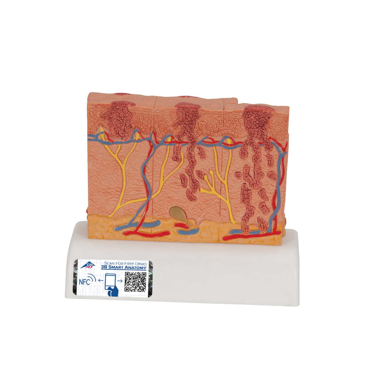
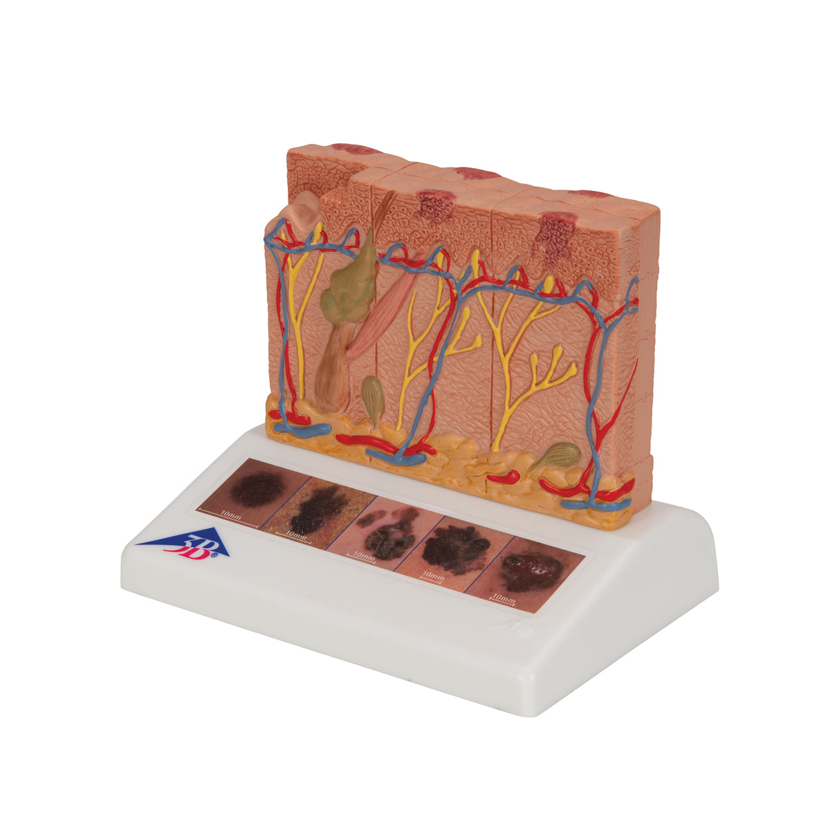
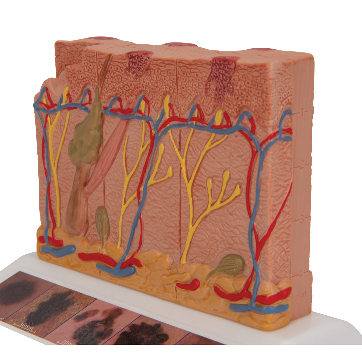
Dermatology and oncology education programs covering melanoma staging and skin pathology benefit from magnified models, and this 8-times 3B Scientific skin cancer model presents five stages of malignant melanoma development alongside healthy skin.
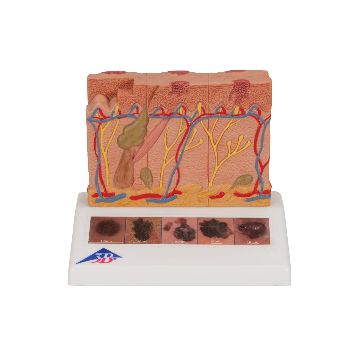
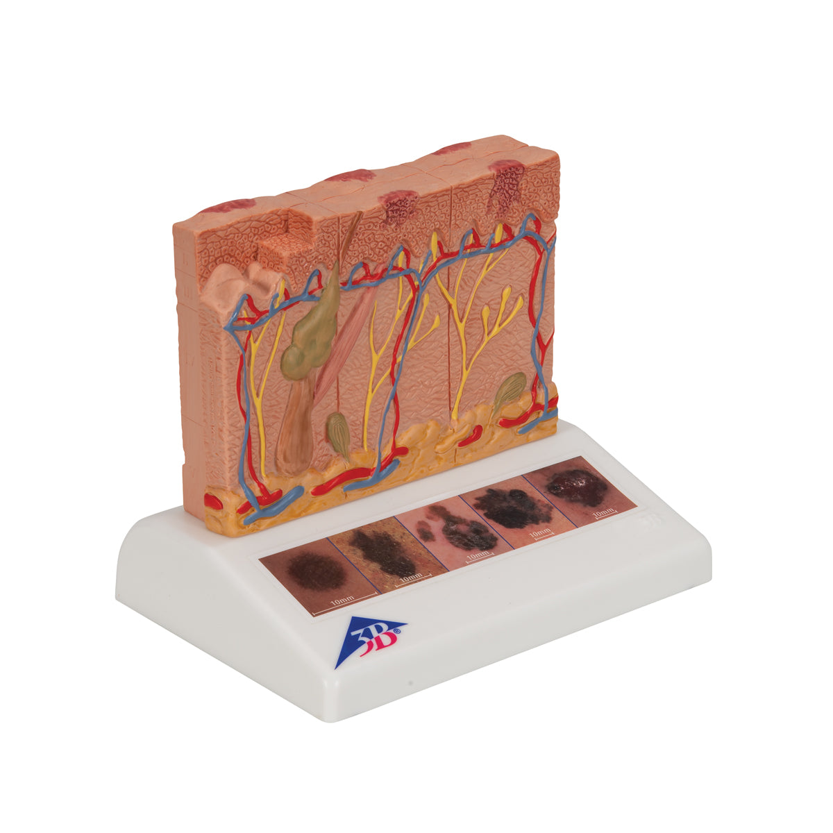
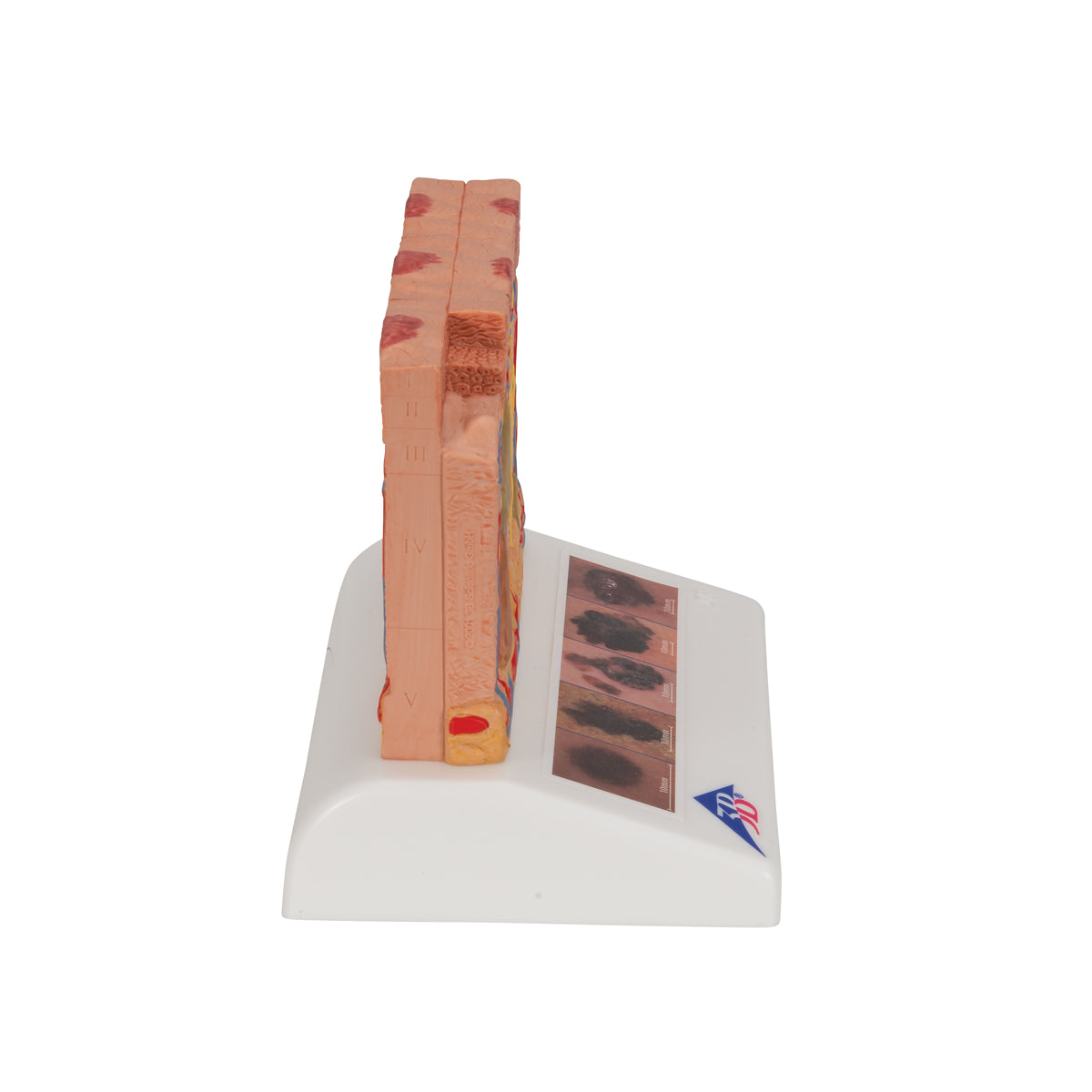
This 3B Scientific® Skin Pathology model shows healthy skin and 5 different stages of malignant melanoma on the front and back, enlarged 8 times:- healthy
- malignant cells are found at the surface, within the epidermis
- malignant cells fill the epidermis, a few invade the papillary layer
- malignant cells fill the papillary layer
- malignant cells invade the reticular layer
- malignant cells have reached the subcutaneous fatty tissue, satellite cells approach a vein
The skin cancer model is a great tool for illustrating this skin pathology.
This product features 3B Smart Anatomy for easy access to model information — read more below.
Specifications
| Weight | 0.34 kg (0.7 lbs) |
| Dimensions | 14 x 10 x 11.5 cm (5-1/2" x 3-7/8" x 4-1/2") |
This item is enhanced with SmartAnatomy!
The new way to experience and supplment your model with online, interactive content. No more reference guides and booklets - access product details right from your mobile device. Learn more

3B Scientific Authorized Distributor

Produced by the 3B Scientific, leaders of high quality healthcare and scientific models. Learn about their story.
Products are backed by the manufacturer warranty.