


Description
Clinical History
A 54-year-old male patient presents with flank pain. He is an active intravenous drug user. Further questioning reveals a history of intermittent haematuria, fevers, malaise, and vomiting. On examination, he is hypertensive and pyrexic. Inspection of his limbs reveals Janeway lesions on his extremities and track marks from recent IV drug use. A systolic murmur is found on auscultation of his chest. Blood tests reveal elevated inflammatory markers, impaired renal function, elevated LDH, and multiple bacteraemic blood cultures. Echocardiogram shows a large mobile tricuspid vegetation. He was commenced on treatment for infective endocarditis but later died from a sudden cardiac arrest.
Pathology
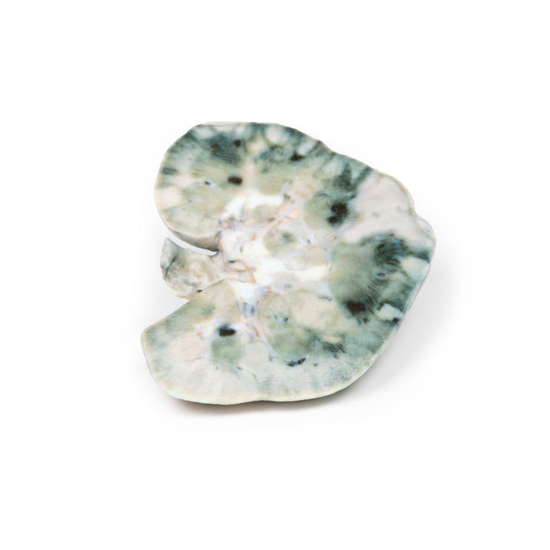
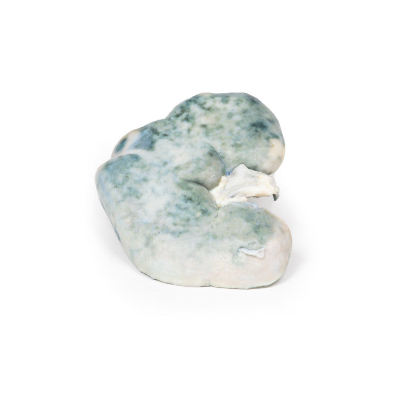
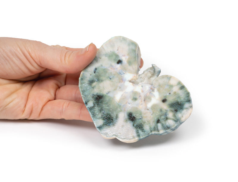
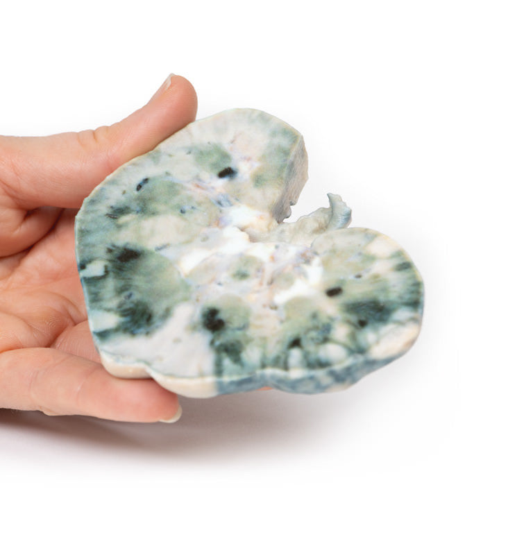
The specimen is the patient’s kidney from post-mortem examination. The kidney has been bisected with a cut half surface on display. There are multiple well-demarcated wedge-shaped pale yellow-white areas evident within the cortex. The base of these pyramids lies against the cortical surface and extends along the renal columns with the apex pointing toward the medulla. The largest is evident at the lateral upper pole of the kidney. These pale areas are infarcted renal tissue. There are dark irregular shaped areas which represent areas of hemorrhage.
Further Information
Renal infarction results from an interruption in the blood flow to the kidney. The kidneys receive almost a quarter of the cardiac output but have limited collateral circulation. The cortex is the most susceptible area to infarction given the blood supply is from proximal to distal. The main causes of interruption of this circulation are cardioembolic disease, renal artery damage, hypercoagulable states, or idiopathic.
Cardioembolic causes are the most common. These include post-myocardial infarction mural thrombi, septic emboli from infective endocarditis, and emboli from mechanical valves. Idiopathic renal infarction is the second most common cause. Damage to the renal artery is the third most frequent cause and includes renal artery dissection, acute vasculitis of polyarteritis nodosa, trauma, or post endovascular intervention. Hypercoagulable states are the rarest cause of renal infarcts, such as hereditary thrombophilia and antiphospholipid syndrome. Infarction is bilateral in approximately 15% of cases.
Presentation of renal infarction depends on the underlying etiology. It can be clinically silent. Common manifestations include costo-vertebral angle pain, haematuria, hypertension due to increased renin release, nausea, vomiting, and sometimes fever.
Laboratory tests used to aid diagnosis include urinalysis for hematuria and serum creatinine levels, which may be elevated especially in bilateral disease. CT abdomen with contrast is the first-choice radiological investigation. A wedge-shaped perfusion defect is the classic finding. Treatment varies depending on the cause of the infarction but generally involves supportive therapy and treatment of the underlying pathology.
