


Description
Clinical History
A 44-year-old man had a skin lesion on his back that grew slowly. At presentation at A&E several years later, he complained of bone pain, and had hepatomegaly and a pleural effusion. He died shortly afterwards.
Pathology
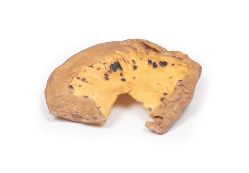
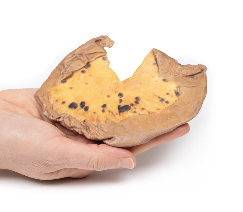
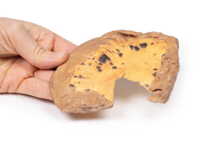
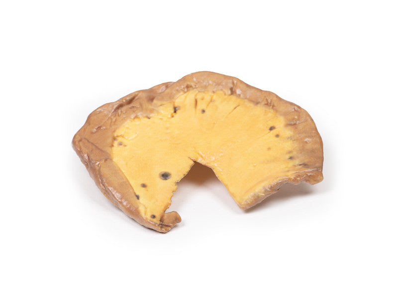
The specimen is a loop of small intestine mounted to display the mesentery, which contains numerous small dark brown, circumscribed nodules varying from pin head size to approximately 1 cm in diameter. Histology confirmed the diagnosis of metastatic melanoma.
Further Information
The most common form of melanoma is cutaneous melanoma, which develops from the pigment-producing cells known as melanocytes. In women, they most commonly occur on the legs, while in men they most commonly occur on the back. About 25% of melanomas develop from moles. Changes in a mole that can indicate melanoma include an increase in size, irregular edges, change in colour, itchiness or skin ulceration.
Skin melanoma is associated with exposure to UV radiation in sunlight or tanning beds. Other risk factors for developing melanoma include fair complexion, presence of large number of melanocytic naevi (moles), severe sunburn as a child, and immunosuppression. It accounts for around 5% of all skin cancer diagnosis but has the highest mortality rate of all skin cancers. Melanomas typically occur in sun-exposed areas as a pigmented lesion with irregular borders, variegated colour, an asymmetrical shape and which evolves with time.
There are multiple mutations common in melanoma. Loss of cell cycle control gene from mutation in CDKN2A gene. Mutations in pro-growth signalling pathways, such as BRAF and PI3K mutations, are seen frequently in melanomas as well as mutations that activate telomerase, such as the TERT gene. Recognition that melanoma antigens activate host immune responses has led to promising immunotherapy, which enhances host T-cell identifying of these antigens.
The most common sites for metastasis of melanoma are the lungs, liver, brain and bone as well as regional lymph nodes, and is highly dependent on the site of the primary tumour. Metastatic melanoma involving the gastrointestinal tract may present with anaemia, overt bleeding, pain, obstruction, or intussusception. The jejunum and ileum are the most commonly involved sites, followed by the colon, rectum, and stomach. Surgery has usually been reserved for patients with the above complications.
The probability of metastatic spread from skin melanoma depends on the stage of the primary tumour, which is based on tumour depth, mitotic activity and ulceration of the skin as well as node and solid organ involvement. Diagnosis of melanoma is made with excisional biopsy. Investigation for bone metastasis is done using blood tests (raised Alkaline phosphatase, calcium and LDH), and radiological investigations most commonly X-ray and CT but MRI and PET scans may also be used. Treatment depends on the stage or the tumour as well as the genetic and immune profiles of the melanoma. Treatment usually involves surgical resection, chemotherapy, targeted therapies (e.g., BRAF inhibitors), immunotherapy, radiotherapy or more commonly a combination of treatments.
