

Description
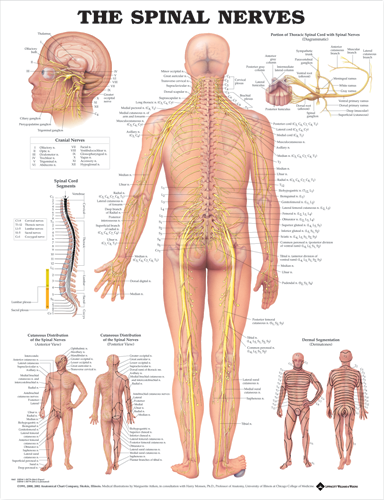
The Spinal Nerves chart maps how the spinal nerves exit the vertebral column and travel through the body, with each nerve labeled against its vertebra. It also covers the cranial nerves and dermatomes, making it a clear reference for teaching and for explaining nerve pathways to patients.
A very popular and useful chart, The Spinal Nerves illustrates the spinal nerves and pathways through the body.
The central illustration shows a posterior view of the spinal nerves exiting from the vertebral column and running throughout the body.
Important skeletal structures are included. All nerves and their corresponding vertebra are clearly labeled.
Also includes:
- detailed illustration of the cranial nerves
- diagrams the portion of the thoracic spinal cord with spinal nerves
- spinal cord segments
- anterior and posterior cutaneous distribution of spinal nerves
- dermal segmentation (dermatomes)
The stone under the foot illustrates the changes in the nerves of a flexed foot.
Dimensions for this chart are 20" x 26" (51cm x 66cm)
How dimensions are measured
Width is measured side to side and height top to bottom, as shown below.
