


Description
Clinical History
A 60-year-old man who had worked in a paint factory for 40 years developed painless haematuria for one month. CT scan showed a suspected tumour in the left renal pelvis. He underwent a nephrectomy.
Pathology
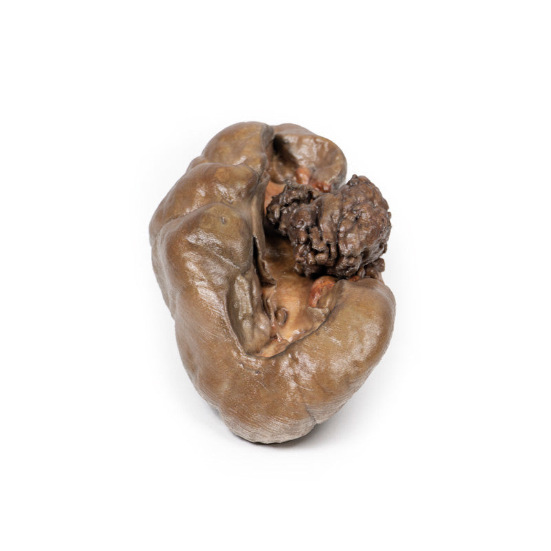
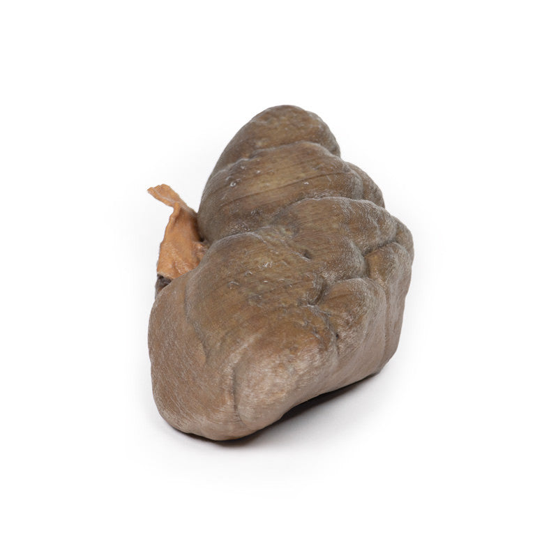
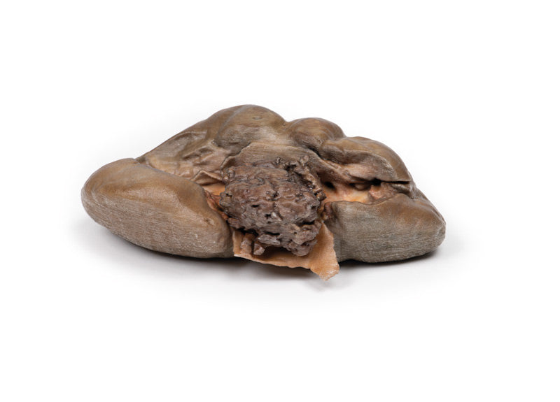
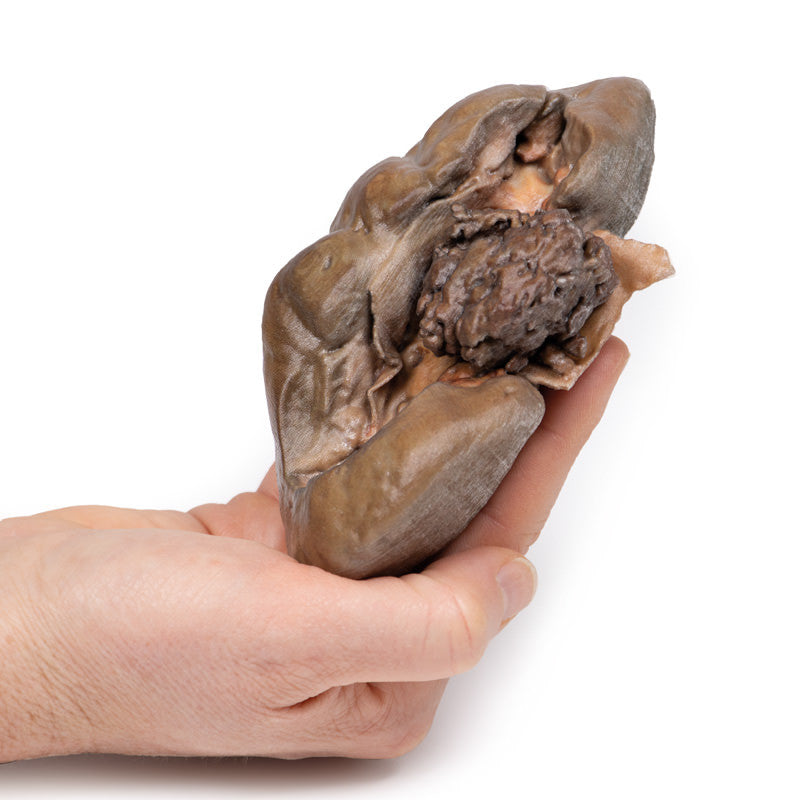
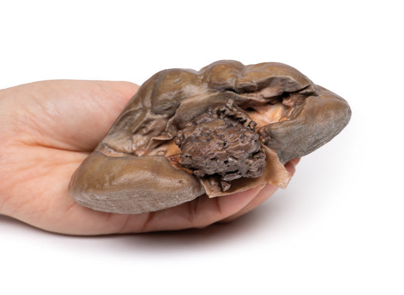
This is the post-nephrectomy kidney. Of note, the kidney maintains its foetal lobulation. There is a friable papillary tumour of 35 mm in diameter projecting into the renal pelvis. The renal pelvis is visibly dilated due to this obstructing tumour. Histological examination revealed this is a papillary transitional cell carcinoma arising in the renal pelvis.
Further Information
Between 5-10% of primary renal cancers arise in the urothelium lining the renal pelvis and calyces. These are similar to tumours which may arise in the ureter and urinary bladder. These tumours range from benign papillomas (rare) to well-differentiated papillary carcinomas, which are common, and poorly differentiated tumours which can be either papillary or flat and infiltrating.
Symptoms of these renal pelvis tumours tend to occur early. Due to the friable nature of the tumours, haematuria is common. As the tumours grow, obstructive symptoms such as palpable hydronephrosis and flank pain can be noticed. The tumours can sometimes be multiple, involving the pelvis, ureter, and bladder.
There is an increased risk of developing urothelial tumours in individuals with Lynch syndrome and analgesic nephropathy. Smoking significantly increases the risk of developing urothelial tumours. Industrial chemicals called aromatic amines, such as benzidine and beta-naphthylamine, which are sometimes used in the dye industry, can lead to urothelial cancers.
Infiltration of the wall of the pelvis and calyces is common in these tumours. The prognosis with infiltration is not good. Five-year survival rates vary from 50-100% for low-grade and non-invasive lesions to 10% for high-grade infiltrating tumours.
