


Description
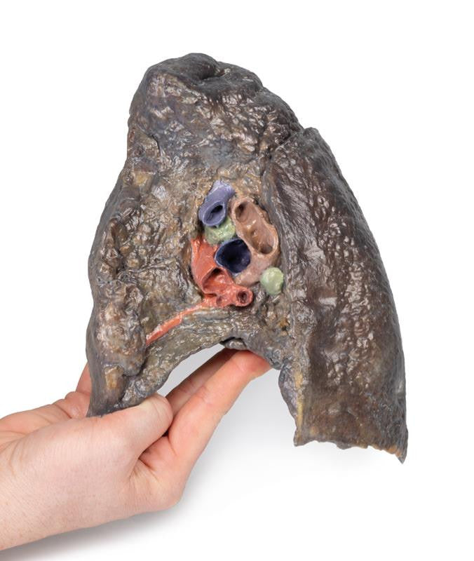
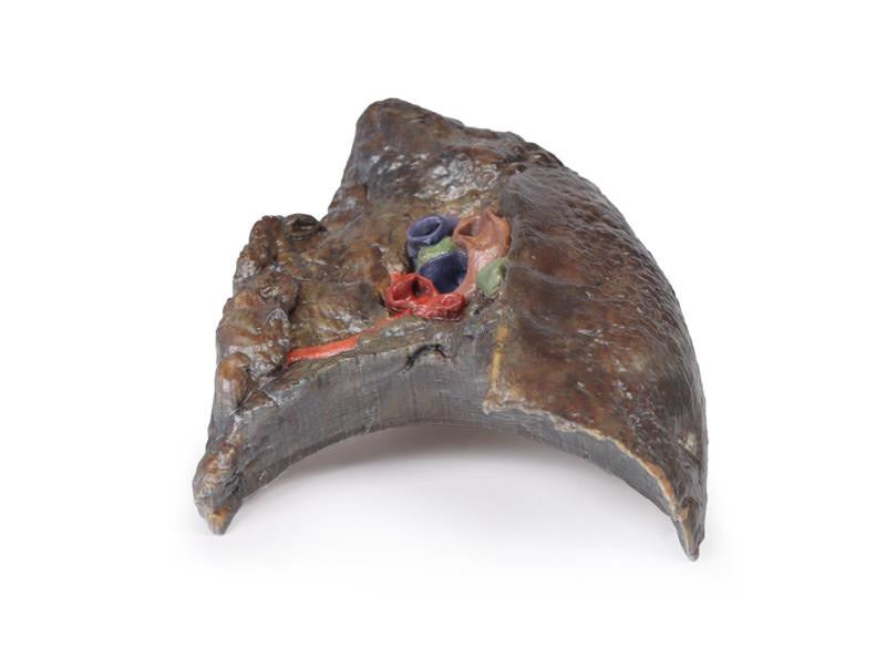
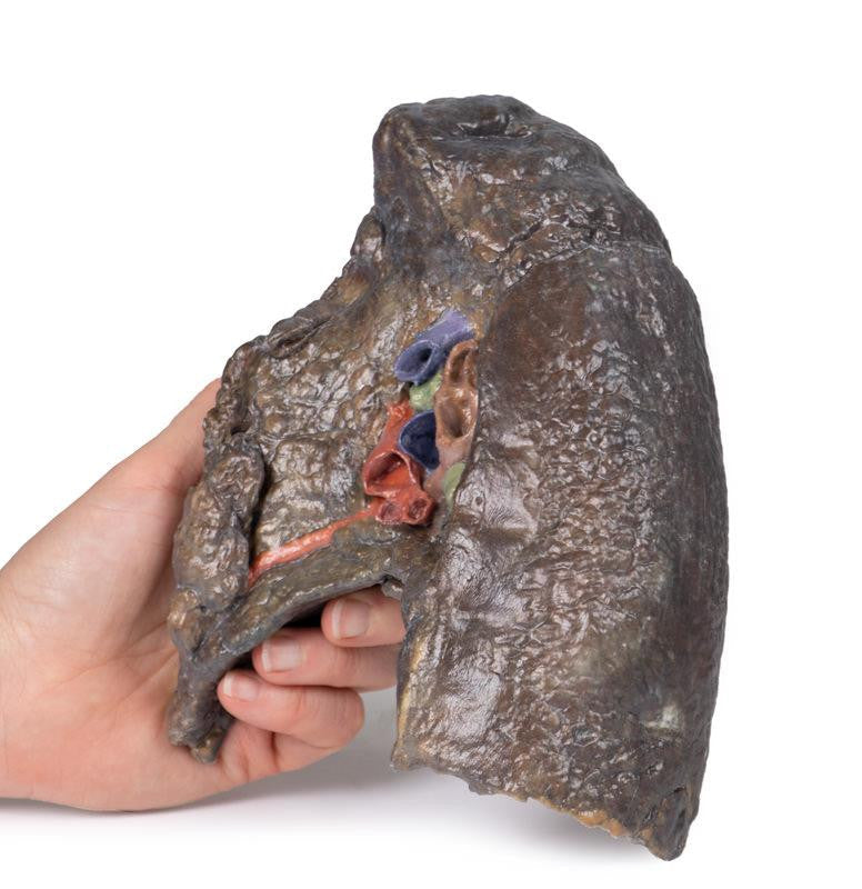
Hilum of the Right Lung
The hilum of a lung is the point at which visceral and parietal pleura meet and functions with the pulmonary ligament as the lung's only connection with the rest of the body. This connection includes the pulmonary artery, superior and inferior pulmonary veins, main bronchi, nerves, and lymphatics.
As the definition of an artery involves carrying blood away from the heart, this will be deoxygenated blood in the pulmonary system, in contrast with the systemic circulation. Similarly, veins carry blood towards the heart, meaning it will be oxygenated in the pulmonary system.
With the specimen cut in a sagittal plane in line with the cardiac impression, nerves and lymphatics are difficult to identify; however, the groove from the oesophagus as it descends posteriorly to pierce the diaphragm can be seen alongside the cardiac impression (of the right atrium), which is notable anterior to the hilum of the right lung. The right main bronchus and its subsequent divisions into lobar bronchi are found more posterior in the hilum; the pulmonary artery and its divisions are located most superior within the hilum; the superior and inferior pulmonary veins and their divisions are most inferior and anterior in the specimen. The oblique and horizontal fissures run along the lateral surface of the specimen, and the hilar lymph nodes are located around the hilum on the medial surface of the lung.
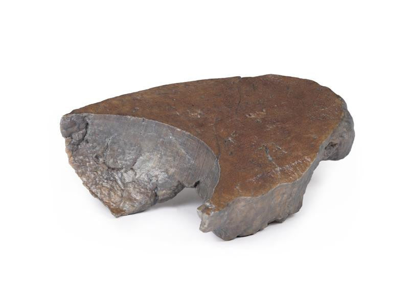
The diaphragmatic surface is found inferiorly, and the costal visceral surface is on the posterior aspect of the specimen.
