


Description
Buy two of our most popular models as a bundle and save. These models show some of the most detailed views of the entire structure including skeletal, muscular, vascular, nervous and connective features.
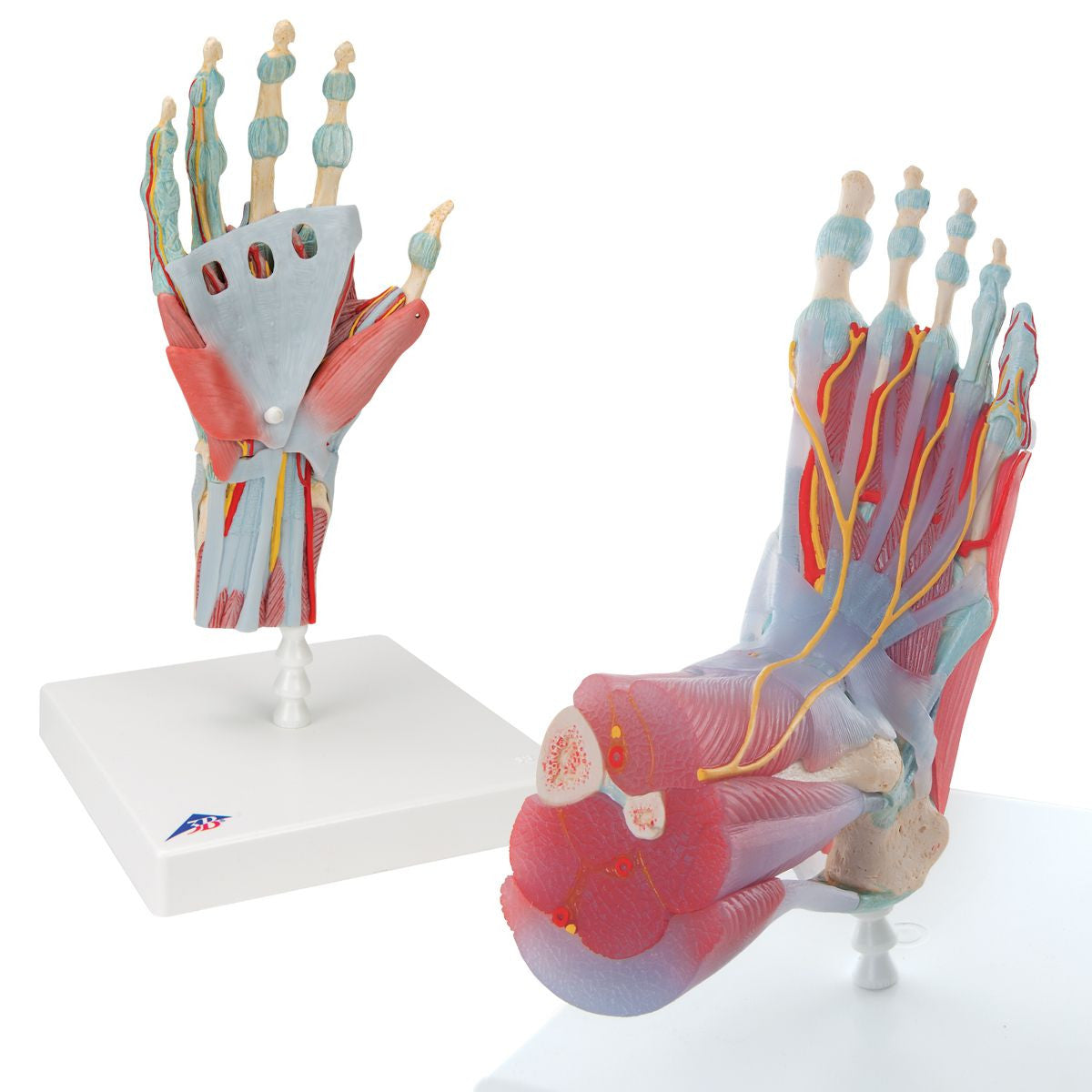
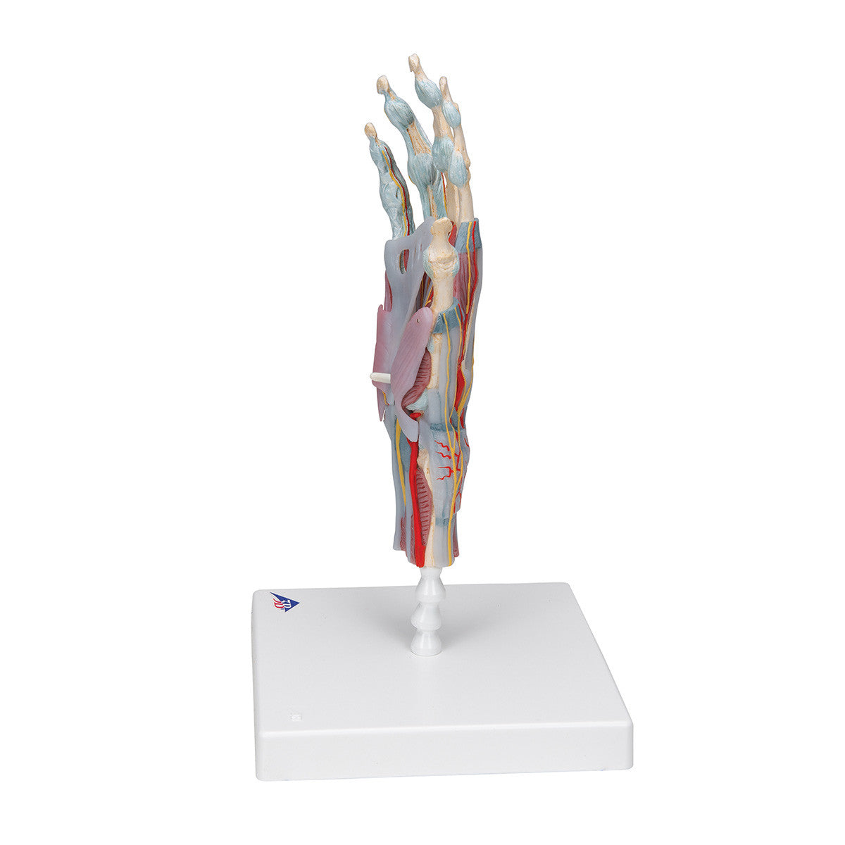
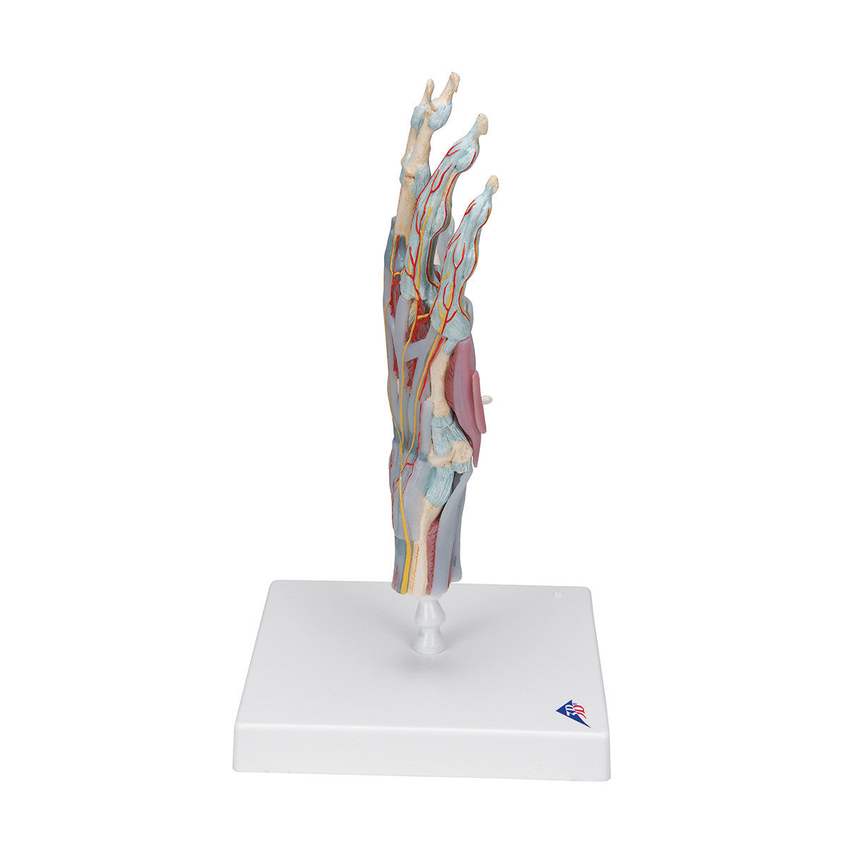
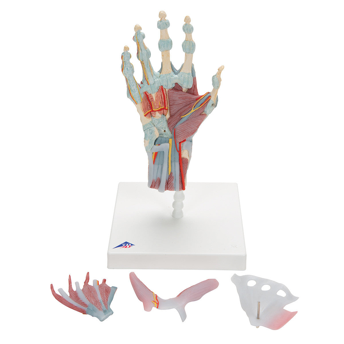
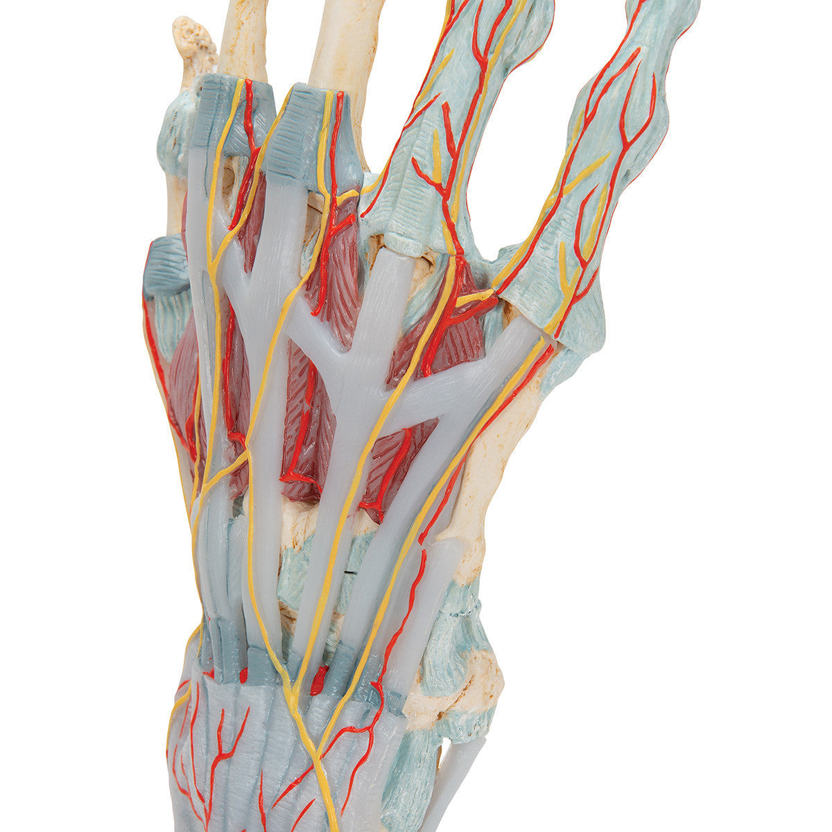
Hand Skeleton Model with Ligaments and Muscles, 4 parts
A highly detailed model of the human hand which allows study of layers of muscles, tendons, nerves and ligaments. This model can be separated into 4 parts to allow you to view deeper layers.
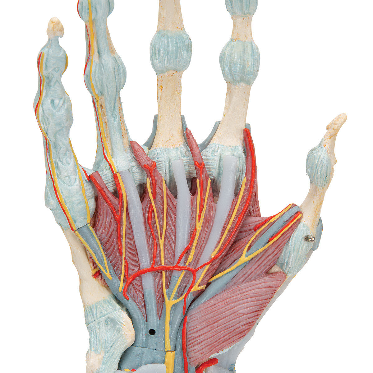
The bones, muscles, tendons, ligaments, nerves, arteries, and veins are all featured in this high quality 4 part model of the hand and lower forearm.
The dorsal side of the hand shows the extensor muscles as well as portions of the tendons at the wrist as they pass under the extensor retunaculum.
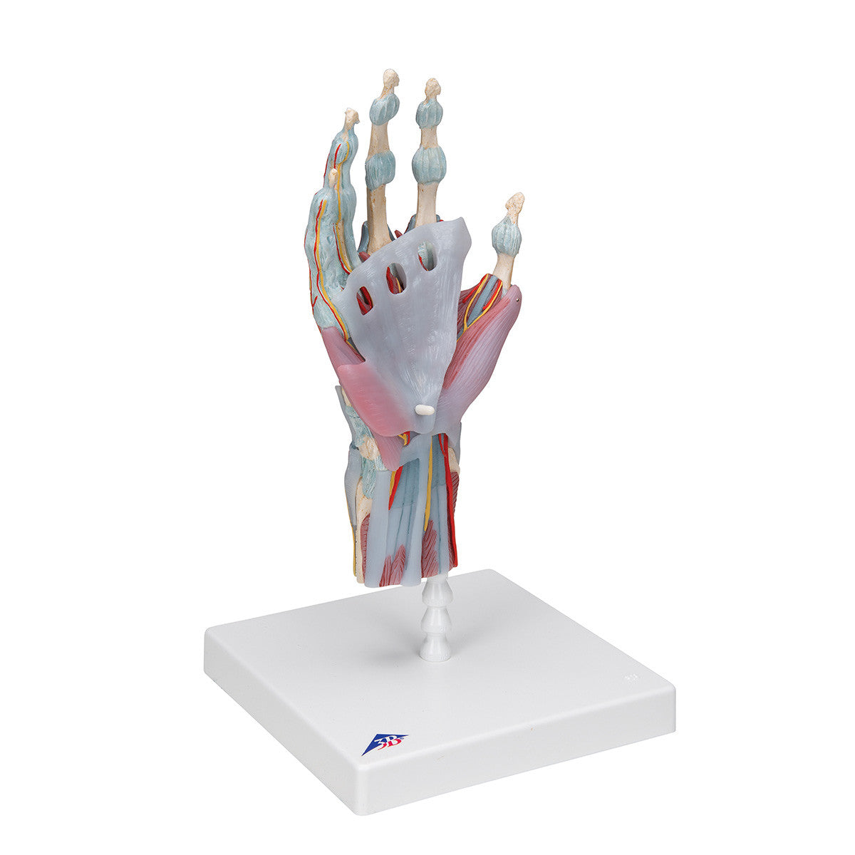
The palmar face of the hand is represented in three layers, the first two are removable to allow detailed study of the deeper anatomical layer of the hand.
In addition clinically important structures such as the median nerve and superficial palmar arterial arch can be explored in detail in the hand model. The deepest anatomical layer allows for study of the intrinsic muscles and deep palmar arterial arch in addition to other details of the anatomy of the hand. This high quality anatomically correct hand model with ligaments and muscles is great for detailed study.
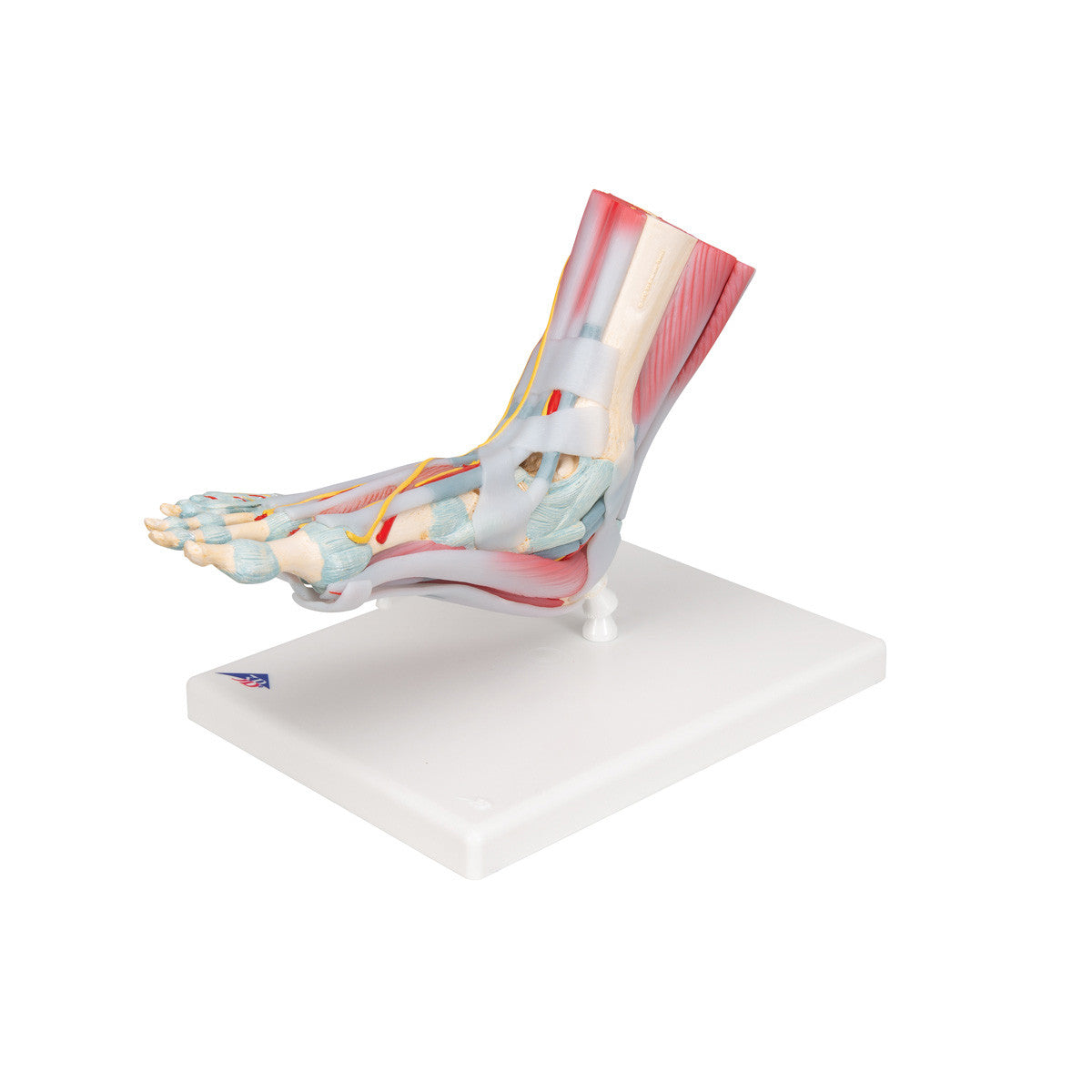
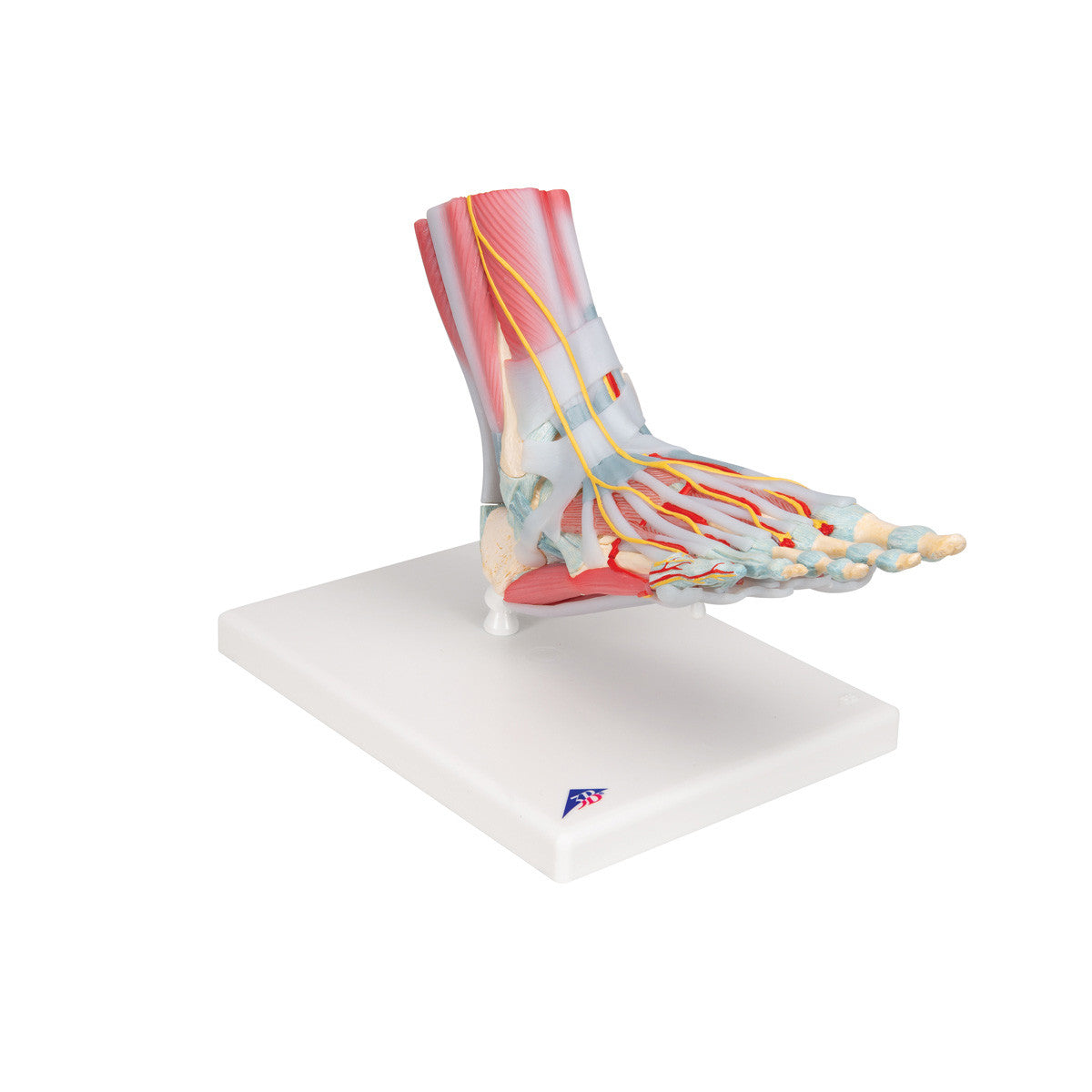
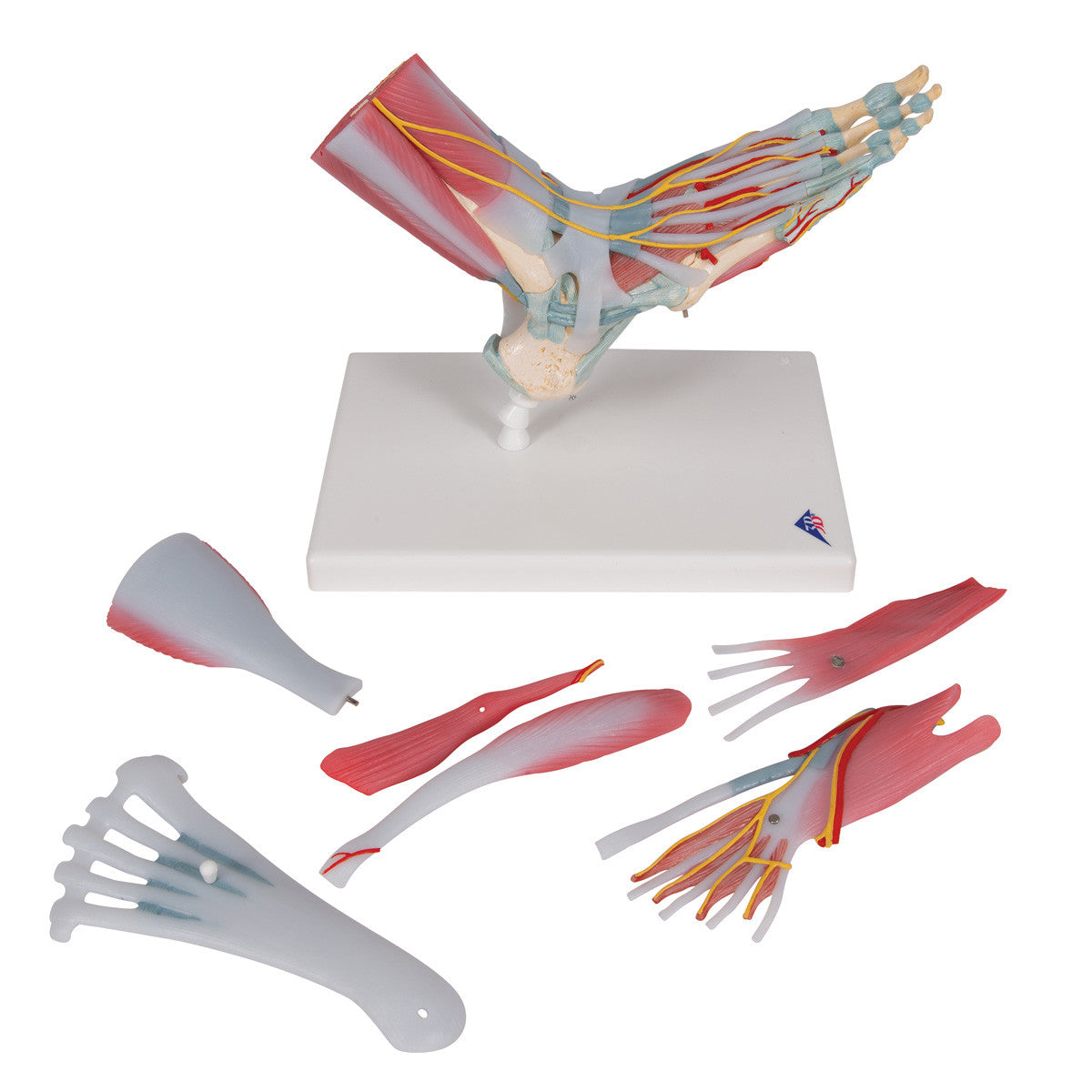
Foot Skeleton with Muscles and Ligaments
All bones, muscles, tendons, ligaments, nerves, arteries, and veins are featured:
- Dorsal side: extensor muscles and portions of the tendons at the wrist passing under the extensor retinaculum
- Palmar face is represented in three layers, removable for detailed study of the deeper anatomical layer (median nerve, superficial palmar arch)
- Deepest anatomical layer shows the intrinsic muscles and deep palmar arterial arch
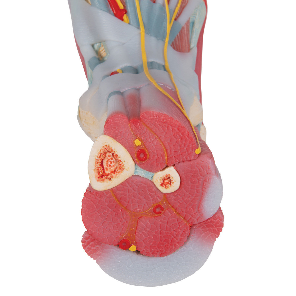
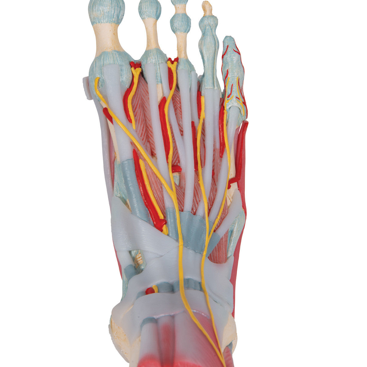
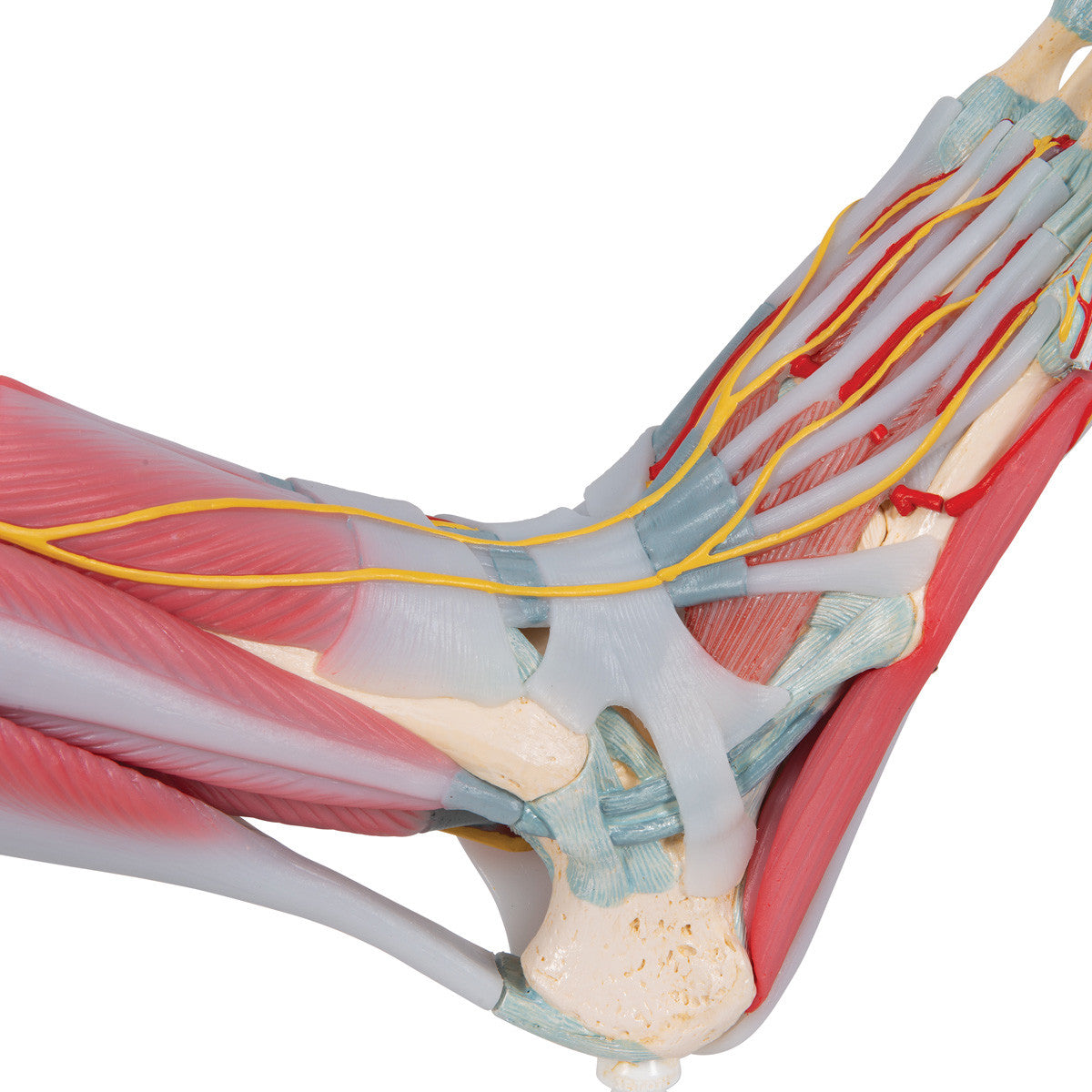
This anatomically detailed model of the foot and lower leg can be disassembled into 6 removable parts for detailed study of the foot and ankle. The foot skeleton features not only the bones but also the muscles, tendons, ligaments, nerves, arteries, and veins of the foot.
The frontal view of the foot model features the extensor muscles of the lower leg. The tendons can be followed on their passage under the transverse and crucial crural ligaments all the way to their insertion points. In addition all tendon sheaths of the foot area are visible. On the dorsal portion of the foot the gastrocnemius muscle is removable to reveal deeper anatomical elements.
The sole of the foot is represented in three layers; the first layer displaying the flexor digitorum brevis. This muscle can be removed from the foot revealing the quadratus plantae, the tendon of the flexor digitorum longus, and the flexor hallucis muscle. This second layer is in turn removable to display even deeper anatomical details of the foot. This foot skeleton model with ligaments and muscles is the best of its kind in quality and value.
3B Scientific Authorized Distributor

Produced by the 3B Scientific, leaders of high quality healthcare and scientific models. Learn about their story.
Products are backed by the manufacturer warranty.