


Description
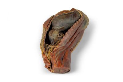
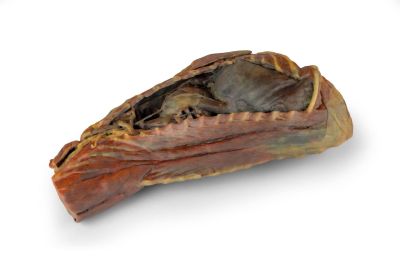
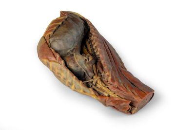
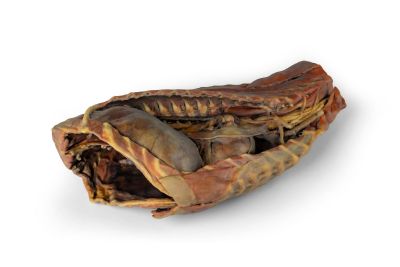
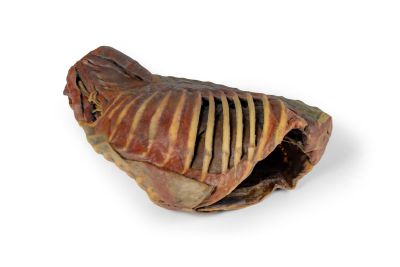
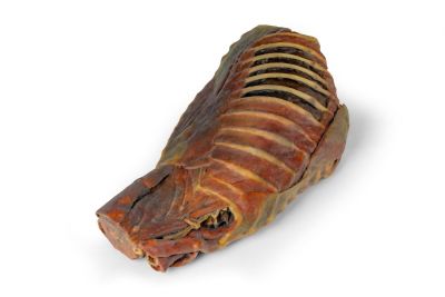
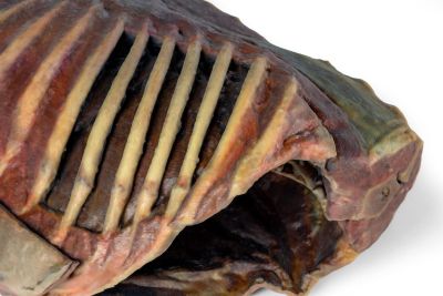
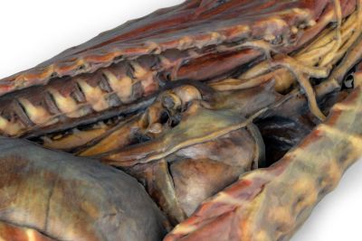
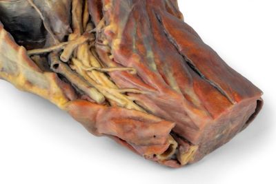
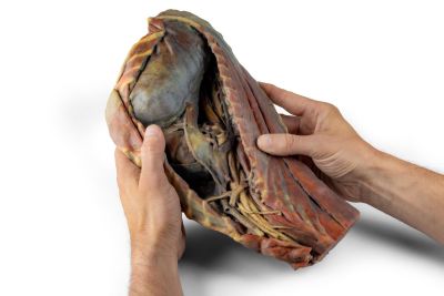
This specimen presents a right-side dissection of the thoracic cavity (Cavitas thoracis) of an adult dog. Following removal of the right thoracic wall, including the ribs (costae), the right lung (pulmo dexter) has been excised, leaving the pulmonary vessels (vasa pulmonalia) in situ in relation to the heart (cor). In the cranial mediastinum (mediastinum craniale), the veins forming the cranial vena cava (vena cava cranialis) are identifiable, along with the course of branches from the right subclavian artery (arteria subclavia dextra). Also visible in this region are the thoracic portion of the longus colli muscle (musculus longus colli), the thoracic trachea (trachea thoracica), and the thoracic esophagus (esophagus thoracicus).
The path of the phrenic nerve (nervus phrenicus) and the vagosympathetic trunk (truncus vagosympathicus) is evident, as is the location of the stellate ganglion (ganglion stellatum). In the middle mediastinum (mediastinum medium), the heart (cor), partially covered by the fibrous pericardium (pericardium fibrosum), is visible. At its base (basis cordis), the pulmonary vessels and the principal bronchi (bronchi principales) — formed by the division of the trachea — can be observed.
The caudal mediastinum (mediastinum caudale) reveals the course of the caudal vena cava (vena cava caudalis) and the right phrenic nerve (nervus phrenicus dexter), adjacent to the caudal mediastinal recess (recessus mediastini caudalis), which is defined by the fold of the caudal vena cava (plica venae cavae). The dorsal aspect is occupied by the esophagus (esophagus), along which travel the dorsal and ventral vagal trunks (truncus vagalis dorsalis et ventralis), as well as the thoracic aorta (aorta thoracica), the right azygos vein (vena azygos dextra), and the sympathetic trunk (truncus sympathicus).
Caudally, the diaphragm (diaphragma) has been preserved, showing its attachments to the costal arches (arcus costales) and lumbar vertebrae (vertebrae lumbales). Additionally, the extensor musculature of the cervical and thoracic spine (musculi extensores columnae vertebralis cervicalis et thoracicae), the intercostal muscles (musculi intercostales), and the brachial plexus (plexus brachialis) are retained.
