


Description
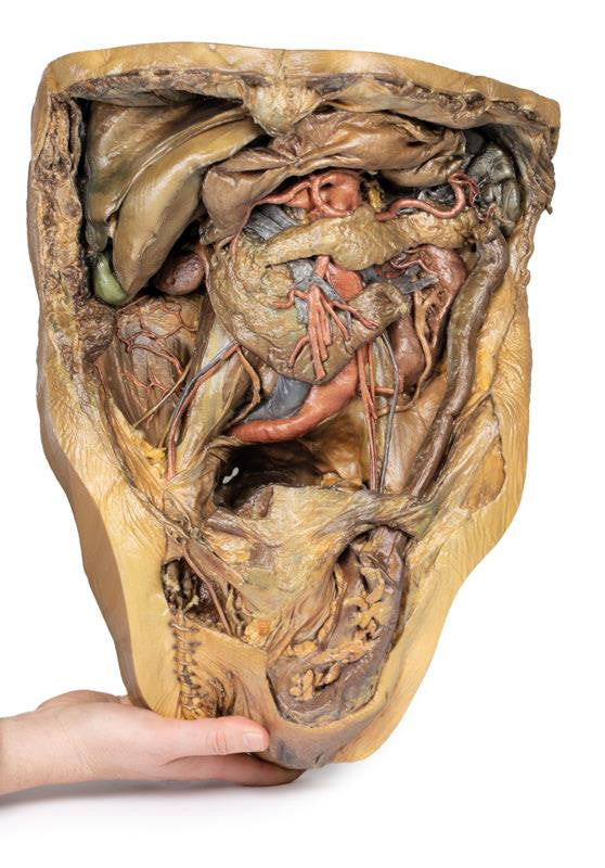
Diaphragm and Xyphoid Process
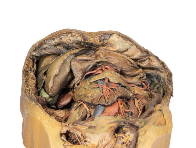
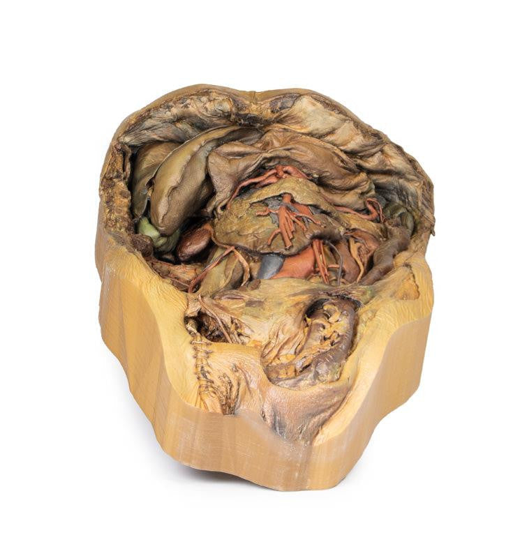
The diaphragm has been secured to the superior border of the dissected specimen with sutures to ensure an unobstructed view of the abdomen. The xyphoid process is in the middle of this sutured border.
Liver and Gallbladder
The liver in the right hypochondrium is pushed laterally to reveal the kidney behind it. The falciform ligament divides the liver’s right and left lobes and contains the ligamentum teres, a remnant of the fetal umbilical vein. Below the ligamentum teres, the gallbladder sits between the liver lobes.
Stomach and Splenic Vasculature
The deflated stomach is pushed up to reveal the twisted splenic artery and vein, which branch into the spleen’s hilum.
Spleen and Pancreas
The spleen sits in the left hypochondrium, with its gastric impression marking the stomach’s greater curvature. The pancreas tail, intraperitoneal and fused to the spleen’s hilum, lies near its inferior pole.
Kidneys
The kidneys are mostly retroperitoneal, but the covering peritoneum is removed in this specimen. Normally, the right kidney lies lower due to the liver, but here it is smaller and higher than the left. The left kidney is enlarged, with two accessory renal arteries from the aorta supplying its hilum and lower pole.
Adrenal Glands
The left adrenal gland is detached from its usual position on the superior pole of the kidney. The middle adrenal artery originates directly from the aorta, left of the coeliac trunk, whilst the inferior adrenal artery is derived from the left renal artery: both supply the adrenal gland. The superior adrenal artery has been obscured by connective tissue.
Rectum and Bladder
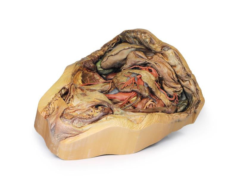
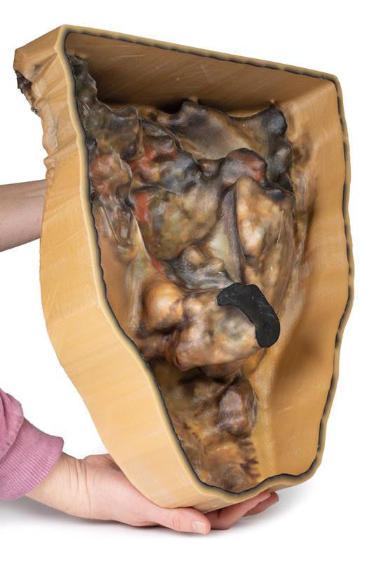
Although the majority of the peritoneum in the abdomen has been removed below the level of the sacral prominence (S1), a layer of peritoneum remains intact, which overlays the rectum and bladder. Notably this the first part of the rectum, which is intraperitoneal.
Gastrointestinal Tract
The final part of the ascending duodenum and the descending colon at the left colic flexure has been ligated with twine, with the intestine in between being removed in order to provide a better view of the abdomen.
Pelvic Region
In this specimen, the sigmoid colon has herniated indirectly through the inguinal canal. On the right, the vas deferens exits the superficial inguinal ring toward the right testicle, while other spermatic cord contents are removed. Sutures below the vas deferens mark the embalming entry via the right femoral artery.
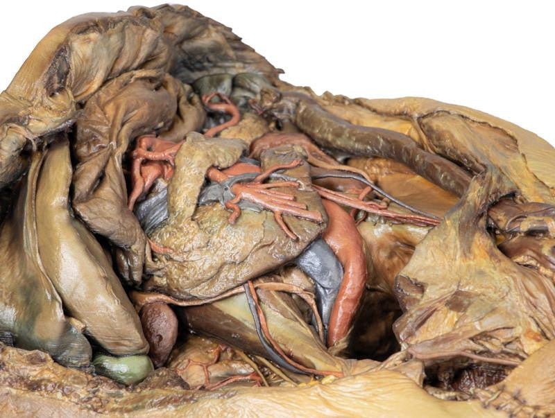
Abdominal Vasculature
The coeliac trunk, located just below the stomach, typically branches into the left gastric, splenic, and common hepatic arteries to supply the foregut.
In this 3D model, the coeliac trunk gives off right and left gastric arteries, the splenic artery, and a gastroduodenal branch that splits into two superior pancreaticoduodenal arteries. The proper hepatic artery arises independently from the abdominal aorta and gives off the right inferior phrenic artery. The iliolumbar artery emerges deep to the right psoas, connecting with branches of the right deep circumflex iliac artery along the iliac crest.
